Antigen-specific cytotoxic T lymphocytes (CTL) kill target cells by recognizing the antigen peptides bound to major histocompatibility antigen (MHC)-I molecules on the cell surface. It is one of the body’s important immune defense responses for anti-tumor, anti-graft and effective control of various infections. The MHC peptide tetramer composed of human leukocyte antigen (HLA) heavy chain and β2 microglobulin light chain, antigen peptide and dye-labeled connexin, has the characteristics of high sensitivity, specificity and efficiency. By using MHC-peptide tetramers, we can not only directly count antigen-specific T cells, but also analyze their phenotype and function, which greatly improves our understanding of the role of antigen-specific CTL in diseases.
MHC-Peptide Tetramer
As early as 1937, Peter discovered the first histocompatibility antigen and led to the discovery of the H-2 histocompatibility antigen complex in mice. Later, a similar complex was found in the human body and named it human leukocyte antigen (HLA). These complexes in other animals are called major histocompatibility complexes (MHC). T cells recognize short peptides presented by MHC molecules through specific TCRs, so fluorescently labeled MHC-peptide complexes can be used to detect antigen-specific T cells. However, the affinity between TCR and MHC-peptide complex is very low, extremely unstable, and the dissociation time is less than 1 min[1]. The use of MHC-peptide complex multimer can increase the affinity with T cells to solve this problem. The MHC-peptide complex multimer can bind multiple TCRs at the same time, which greatly slows down the dissociation speed, thereby increasing the positive rate of detection. Altman et al. prepared MHC peptide tetramers based on this principle[2]. The light and heavy chains of MHC are produced by Escherichia coli, and they are recombined with antigen peptides by mole in vitro to form MHC-peptide complexes. After purification, a biotin molecule is connected to the C-terminus of the heavy chain of the MHC-peptide complex, then a fluorescein labeled streptavidin with four biotinylated MHC-peptide complex binds to form a tetramer (as shown Fig.1).
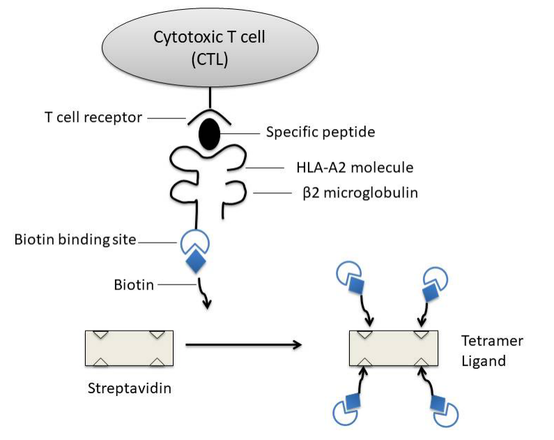
Figure 1: Diagram of MHC peptide tetramer: biotinylated MHC I-antigen peptide complex is mixed with fluorescently labeled avidin to form a tetramer
MHC-peptide tetramer action principle
Major histocompatibility antigens are glycoproteins located on the surface of cells. They are highly polymorphic and are antigen-presenting molecules that play a key role in immune response. The antigens that antigen-specific CTLs can recognize are mainly peptides derived from endogenous proteins and bound to MHC-I molecules. MHC-I molecules are heterodimers composed of a highly polymorphic heavy chain and a conservative light chain, β2 microglobulin [3], which are expressed in all nucleated cells. Antigen short peptides that are usually combined with MHC-I molecules are synthesized in antigen presenting cells (APC) (endogenous antigen) and binds in the peptide binding groove formed by the α1 and α2 functional regions of the heavy chain of MHC-I molecules in the endoplasmic reticulum of the cell. Subsequently, due to the non-covalent binding of β2 microglobulin to the heavy chain, MHC-I peptide complexes are more stable, and they are finally located on the cell surface. By interacting with the αβ T cell receptor (TCR) on the surface of CD8+ antigen-specific CTLs, CTLs are activated to kill infected cells.
Construction of MHC-II peptide tetramer
The basic principle is similar to the construction of MHC-I peptide tetramer. The two heavy chains of the modified MHC-II molecules are synthesized in vitro, biotinylated, and reacted with avidin to form tetramers.
Application of MHC peptide tetramer
- Quantitative analysis of specific T cells (CTLs or Th cells) that recognize a specific antigen
Tetramers are highly specific and sensitive. This feature is particularly important in the detection of certain diseases with a small number of T cells. Such as chronic hepatitis B and C virus infections, a small number of T cells not only make detection difficult, but also affect the understanding of their functions.
- Phenotypic analysis of tetramer positive cells
Combine with flow cytometry to detect multi-fluorescence markers to analyze the expression of its surface molecules, such as CD45 isomeric molecules, activation markers, homing markers CCRS, CCR7, etc., or combined with intracellular staining to detect the expression of some effector molecules or related markers, such as perforin and cell proliferation marker Ki67.
- Isolate the group of cells and perform cell function analysis (combined with other methods)
Used for the secretion and release of a specific cytokine, such as detecting the amount of secreted TNF-α to reflect the killing activity of the CTLs to be tested; or performing a cytotoxicity test to directly observe the killing function of the cell. Although this sorting of cells can also be done by magnetic beads, the use of tetramers is more economical, simple and straightforward.
- Use these cells for disease treatment and vaccine research
- Use MHC peptide tetramer/antigen-specific T cell lysis method to detect the relative activity of antigen-specific T cells
References
1.Corr, Slanetz AE, Boyd LF, Jelonek MT, Khilko S, al-Ramadi BK, Kim YS,Maher SE, Bothwell AL, Margulies DH. T cell receptor-MHC class I peptide interactions:affinity, kinetics, and specificity. Science. 1994 Aug 12;265 (5174):946-9.
2.Altman JD, Moss PA, Goulder PJ, Barouch DH, McHeyzer-Williams MG, Bell JI, McMichael AJ, Davis MM. Phenotypic analysis of antigen-specific T lymphocytes. Science. 1996 Oct 4; 274 (5284):94-6
3.Nepom Gebe J, Wang Buckner Swanson JH, Novak EJ, Reichstetter S, Reijonen H, E, Kwok WW. HLA class II tetramers: tools for direct analysis of antigen-specific CD4′ T cells. Arthritis Rheum. 2002 Jan;46(1):5-12. Review.
]]>

Figure 1 Scanning electron micrograph of a human T cell
In the current mainstream model, T cells identify potential threats through surface “T cell receptors” (TCR). For example, when viruses and bacteria infect the human body, specific proteins on their surfaces are loaded into a structure called the major histocompatibility complex (MHC) for T cells to recognize. In the past, we know a lot about αβ T cell receptors. It is believed that T cell receptors will bind to MHC or MHC-like molecules in a head-to-head manner, thereby recognizing specific signals from pathogens. In this study, the scientists focused on γδ T cell receptors, a relative of these receptors.
In this new study, the researchers describe a cell population in a class of little-known T cells called γδT cells that recognizes an MHC-like molecule called MR1. Using high-intensity X-ray beams on a synchrotron in Australia, they obtained detailed three-dimensional images of the interaction between γδT cell receptors and MR1, and got interesting results: γδT cell receptors bind under MHC-like molecules for recognition. Dr. Jérôme Le Nours, one of the co-first authors of the study, made a very vivid metaphor: “Imagine that (MHC molecule) is like a flag standing on a cell. We always think the t-cells are sitting on top of the flagpole, reading the message. But what we found is that some t-cells bind to the bottom first.”
These are important details that can change our approach to studying T cell biology in the future. This is important because T cells are a key weapon of our immune system, and understanding how T cells target and act on other cells is critical to using their power to develop immunotherapies that treat diseases such as infections and cancer. Researchers believe that this unusual recognition system is expected to help us understand how TCR binds to target molecules. Given that T cells are an important weapon in the body’s fight against disease, these insights are expected to lead to more effective therapies. Because γδ T cells also play a role in fighting cancer, the existing anti-cancer immunotherapy has the opportunity to be further enhanced.
Dr Nicholas Gherardin, co-first author of the paper and the Doherty Institute at the University of Melbourne, said: “Our research indicates that MR1 is a novel molecular target for γδT cells. These cells play a decisive role in the immune response against infection and cancer, but little is known about how they respond. MR1 may signal γδT cells to indicate the presence of viruses or cancer cells and trigger these cells to trigger a protective immune response. ” Researchers will dig deeper into this mechanism and strive to apply it to the treatment of diseases.
References
- Jérôme Le Nours et al. A class of γδ T cell receptors recognize the underside of the antigen-presenting molecule MR1. Science, 2019, doi:10.1126/science.aav3900.

Although MHC class I molecules contribute greatly in alerting the immune system to virally infected cells, it is still not well-understood as to how MHC-1 molecules bind to antigen peptides in the human immune system. Now this puzzle is probably clarified as recently researchers at the University of California, Santa Cruz have unveiled the details of key molecular interactions related to MHC-1 protein selection and processing of antigens. Their new findings, published in the December 3 issue of PNAS.
As is previously known, the role of the MHC-1 protein is to enable every cell in the body to display on its surface fragments of all the proteins that are being produced in that cell. With the help of MHC-1 proteins, cytotoxic T cells can more easily recognize foreign proteins derived from pathogens or mutant proteins produced by tumor tissues and make quick immune responses accordingly.
Sgourakis, assistant professor of chemistry and biochemistry at the University of California, Santa Cruz, and his team worked closely with Erik Procko’s group at the University of Illinois to understand the mechanism of protein-peptide selection and binding to MHC-I proteins. This new paper reveals how the interaction between MHC-1 protein and molecular chaperone affects the composition of displayed antigens.
There are thousands of different variants of the human MHC-1 protein, produced by different “alleles” of the MHC-1 gene. The polymorphism of MHC-1 protein is the cause of many individual differences in the immune response, including differences in sensitivity to autoimmune diseases, infections, and cancer. Each person has six major MHC-1 alleles (three from the mother and three from the father). These six MHC-I proteins can load all the peptides produced in a cell.
Sgourakis’ team studied four different MHC-I alleles and their interactions with molecular partners and antigens. Nuclear magnetic resonance (NMR) technology was used to reveal dynamic structural changes in MHC-1 protein. One of the functions of chaperones is to help MHC-1 proteins fold into their active shapes and stabilize them to prevent misfolding and aggregation. However, only some MHC-1 alleles rely on chaperone proteins for antigen loading. It still remains a mystery as to why some molecules rely on chaperone proteins, while others don’t. The researchers found that if the three-dimensional structure of the MHC-1 molecule is rigid, the chaperones are not involved in antigen loading. However, if it is flexible in the peptide-binding groove, the chaperone will interact with it and help the antigen loading process through ejecting antigens with low affinity to the binding groove, thereby ensuring that the MHC-1 protein binds only high-affinity antigens that can be displayed on the cell surface in the appropriate conformation, and thereby activating the T cell response.
By using flow cytometry and with the advance in fluorescently labeled tetrameric MHC–peptide complex, it has been made possible to directly visualize, quantify and characterize antigen-specific T cells, which has nearly transformed scientists’ understanding of cellular immune responses.
References
- McShan, A. C., Devlin, C. A., Overall, S. A., Park, J., Toor, J. S., Moschidi, D., … & Sgourakis, N. G. (2019). Molecular determinants of chaperone interactions on MHC-I for folding and antigen repertoire selection. Proceedings of the National Academy of Sciences.
Related Products
]]>