What is solid-phase peptide synthesis (SPPS)?
Solid-phase peptide synthesis (SPPS) is an important method for synthesizing peptides. In SPPS, peptides are synthesized on an insoluble resin that serves as the solid support. The process begins by attaching the C-terminal amino acid to the resin, and subsequent amino acids are added step by step in a sequence dictated by the desired peptide. Each elongation cycle typically consists of deprotection (removing a protecting group from the amino acid) and condensation (forming a peptide bond between the activated amino acid and the growing chain).
The most challenging part of peptide synthesis is the formation of the peptide bond, which requires an efficient activation of the carboxyl group to promote its reaction with the amino group. This activation is achieved through the use of condensation agents, which are chosen based on factors such as compatibility with the resin, reaction efficiency, and minimization of side reactions.
General process of synthesis
- First, the initial amino acid (the C-terminal residue) is attached to the resin (loading).
- After the amino acid binds to the resin, the resin is washed to remove by-products and excess reagents.
- Next, the amino protecting group is removed, and the resin is washed again. Under the action of a coupling agent, the next amino acid is coupled to the peptide-resin complex. This coupling and washing cycle is repeated until the entire peptide sequence is completed.
- Finally, all protecting groups are removed, the peptide-resin is washed, and the peptide is cleaved from the resin.
Key Considerations in Choosing a Condensation Agent
The selection of a condensation agent is crucial for ensuring efficient peptide bond formation. Key factors influencing the choice of a condensation agent include:
Activation Efficiency: The reagent must activate the carboxyl group of the amino acid efficiently to form the reactive intermediate, which will then react with the amine group of the next amino acid.
Minimization of Side Reactions: The condensation agent should ideally minimize side reactions such as racemization, hydrolysis, and degradation of sensitive amino acids or functional groups.
Compatibility with the Resin: The reagent must not interfere with the resin or other components of the reaction mixture, including solvents and protecting groups.
Solubility: Condensation agents should be sufficiently soluble in the solvent system used during SPPS to achieve optimal reaction conditions.
“Orthogonal” protection in solid-phase peptide synthesis
Many amino acid side chains are reactive and, if not protected, can form by-products. To successfully synthesize peptides, side chains need to be protected, and these protecting groups must remain stable throughout repeated deprotection of the amino group. Ideally, the amino-protecting group and side-chain-protecting group can be removed under completely different conditions-such as removing the amino-protecting group under basic conditions and the side-chain-protecting group under acidic conditions. This approach is known as “orthogonal” protection. Common combinations include Boc/Bzl protection and Fmoc/tBu protection. In the Boc/Bzl protection scheme, the Boc group temporarily protects the amino group, while the benzyl group provides more permanent protection for side chains. Although both Boc and benzyl protecting groups are acid-sensitive, making Boc/Bzl not a truly orthogonal protection strategy, it is still widely used. This is because the Boc group can be removed under mild acidic conditions (e.g., 50% TFA in DCM solution), whereas the benzyl group requires a very strong acid (such as HF or TFMSA) for removal.
Condensing agents in solid-phase synthesis
Carbodiimides
Carbodiimides, such as dicyclohexylcarbodiimide (DCC) and 1-(3-dimethylaminopropyl)-3-ethylcarbodiimide (EDC), are among the most widely used condensation agents in peptide synthesis. DCC, DIC, EDCI. Dicyclohexylcarbodiimide (DCC) and diisopropylcarbodiimide (DIC) are commonly used to convert carboxylic acids into amides, esters, and anhydrides. They can also convert primary amides into nitriles, which may lead to side reactions when aspartic acid and glutamic acid residues are present in peptides. DCC generates dicyclohexylurea as a by-product, which is nearly insoluble in most organic solvents and precipitates out of the reaction mixture as the reaction progresses. Therefore, DCC is highly useful in solution-phase reactions but less suitable for reactions on resin. In solid-phase synthesis, DIC is used because its urea by-product is more soluble and remains in solution. In some applications, such as protein modification, ethyl-(N’,N’-dimethylaminopropyl) carbodiimide hydrochloride (EDCI) is used. This carbodiimide reagent and its urea by-product are water-soluble, allowing by-products and excess reagent to be removed with water washes.
In peptide synthesis, to prevent racemization of amino acids during carbodiimide activation, an equivalent amount of 1-hydroxybenzotriazole (HOBt) is commonly added along with the carbodiimide.
Mechanism of Action: Carbodiimides activate the carboxyl group of an amino acid by forming an acyl-urea intermediate. This intermediate then reacts with the amino group of another amino acid, resulting in the formation of a peptide bond.
Advantages: Carbodiimides are relatively inexpensive and easy to handle. They can be used for a wide variety of amino acids and peptides, and they are effective under mild conditions, minimizing the risk of side reactions such as racemization.
Limitations: One of the significant drawbacks of carbodiimides is the formation of urea byproducts, such as dicyclohexylurea (DCU) when DCC is used. These byproducts can precipitate out of solution and require removal by filtration, which can reduce the yield. Moreover, carbodiimides can also lead to the formation of side products due to incomplete activation.
Benzotriazole Derivatives
Benzotriazole derivatives, such as 1-hydroxybenzotriazole (HOBt), benzotriazole-1-yl-oxy-tris-pyrrolidino-phosphonium hexafluorophosphate (BOP), and Oxyma, are frequently used in peptide synthesis to improve the efficiency of carbodiimide-based activation.
To avoid racemization and side reactions associated with carbodiimide condensing agents, many alternative reagents have been developed to generate OBt esters in situ. Benzotriazol-1-yloxytris(dimethylamino)phosphonium hexafluorophosphate (BOP) was one of the first reagents developed. BOP does not lead to dehydration of asparagine and glutamine side-chain amides to form nitrile by-products, and it causes minimal racemization of amino acids. However, BOP produces highly carcinogenic hexamethylphosphoramide as a by-product in condensation reactions, so it must be handled carefully. PyBOP offers comparable condensation efficiency to BOP but with a lower risk from its by-products. PyAOP is also an effective condensing agent, especially for coupling between N-methyl-protected amino acids. PyBrOP is often used for coupling sterically hindered amino acids, such as N-methyl amino acids or α,α-dialkylglycines, where other condensing agents are less efficient. BOP-Cl is commonly used to couple Fmoc-α,α-dialkyl amino acids to tritylphenyl resins.
Mechanism of Action: These reagents work by stabilizing the active ester intermediate that is formed during the activation process. HOBt, for example, reacts with the carbodiimide to produce a highly reactive ester intermediate, enhancing the efficiency and reducing side reactions.
Advantages: The addition of HOBt or related compounds can significantly improve the efficiency of peptide bond formation, reduce side reactions, and suppress racemization, especially for sterically hindered or sensitive amino acids.
Limitations: While these reagents are effective, their cost can be higher compared to carbodiimides alone, and their use requires additional care in handling and disposal due to their chemical reactivity.
Uronium Salts
Uronium salts, such as ethyl(2-dimethylamino-ethyl)-phosphonium tetramethylfluorophosphate (EEDQ) and t-butylphosphonium hexafluorophosphate (TBTU), are another class of reagents used in peptide bond formation. These agents activate carboxyl groups by forming reactive phosphonium or uronium species.
Mechanism of Action: Uronium salts generate reactive species that facilitate the coupling of the carboxyl group with the amine group of the incoming amino acid. These species are highly electrophilic, promoting efficient peptide bond formation.
Advantages: Uronium salts are highly effective in promoting fast and efficient peptide bond formation. They are particularly useful for synthesizing peptides with low solubility or challenging sequences. They also tend to produce fewer side products.
Limitations: Despite their efficiency, uronium salts can be expensive and may lead to contamination with byproducts if not carefully controlled. Their use in SPPS may also be limited by the compatibility with the resin or other reagents.
Phosgene Derivatives
Phosgene derivatives, such as chlorophosgene and triphosgene, are potent agents used for activating carboxyl groups in peptide synthesis.
Mechanism of Action: Phosgene derivatives react with the carboxyl group of amino acids to form an acylated intermediate. This intermediate then reacts with the amine group of the next amino acid, facilitating the peptide bond formation.
Advantages: Phosgene derivatives are highly efficient in peptide bond formation and can work well with challenging amino acids. They offer high yields and can help minimize racemization and side reactions.
Limitations: The major disadvantage of phosgene derivatives is their toxicity and safety concerns, as phosgene is a highly hazardous substance. Additionally, the reagents can be expensive, and their use requires specialized equipment for handling and disposal.
Activation with Mixed Anhydrides
Mixed anhydrides are formed by reacting an amino acid with an acid chloride or an acylating agent. This reaction produces an acylated amino acid intermediate that is then used for peptide bond formation.
Mechanism of Action: The mixed anhydride is a reactive intermediate that can readily react with the amino group of another amino acid, resulting in the formation of a peptide bond.
Advantages: Mixed anhydrides can be highly effective, especially when used in combination with reagents like HOBt or pyridine. The method is versatile and applicable to a broad range of amino acids.
Limitations: One challenge with this approach is that mixed anhydrides are highly reactive, which can sometimes lead to side reactions if the reaction conditions are not carefully controlled. Additionally, the preparation of mixed anhydrides can be more complex than other methods.
New Developments in Condensation Agents
Recent advances in peptide synthesis have led to the development of newer, more efficient condensation agents. These agents aim to address the limitations of traditional reagents by enhancing reaction specificity, improving yields, and reducing side reactions.
One promising area is the development of green reagents, which are designed to be environmentally friendly and non-toxic. For example, Oxyma Pure, a derivative of HOBt, is one such reagent that has gained popularity due to its improved efficiency and lower toxicity profile.
Additionally, solid-phase activated reagents (SPARs) are being explored as a way to streamline the synthesis process by coupling the activation step directly to the solid-phase resin. These reagents eliminate the need for liquid-phase activation, which can reduce solvent consumption and simplify the overall process.
]]>Peptide drugs, as a type of pharmaceutical with molecular weight between small molecule chemical drugs and biopolymers, provide more options for medicinal chemists and have become a hot research topic in recent years. This article discusses the development of analysis methods for peptide-related impurities in peptide drugs.
Features of peptides
Peptide molecules are compounds formed by the connection of multiple amino acids through peptide bonds (amide bonds), which have the following characteristics:
(1) Peptide drugs have a larger molecular weight. For example, the molecular weight of the peptide star drug somatostatin (semaglutide) is 4114 Da, which is lower than that of biopolymer drugs but several times that of small molecule drugs. The increase in molecular weight leads to some important changes in chromatographic behavior, such as increased sensitivity to solvent strength, even slight changes in B% can cause rapid changes in retention time. Furthermore, diffusion in the mobile phase is slower, leading to a decrease in column efficiency and an increase in peak width.
(2)Peptide molecules have a tertiary structure. Peptide chains form secondary and tertiary structures through α helices and β folds, with their unique three-dimensional structure. Due to the molecular volume and three-dimensional structure, not all amino acids can simultaneously contact a fixed phase, only residues on the surface can interact with each other.
(3)Peptide molecules are connected through amide bonds, which have partial double-bond properties. The isomers of cis-trans exchange may cause peak distortion.
(4)Peptide molecules are amphipathic, with acidic carboxyl groups and basic amino groups, and have a specific isoelectric point.
(5)The mass spectrometry of peptide drugs is extremely complex. There are various types of process impurities and degradation impurities in synthetic peptide molecules, especially the structure and properties of racemic impurities are very similar to the main component, and their chromatographic behavior is also similar, which greatly increases the difficulty of separation and analysis.
Analysis methods for peptide-related impurities
Regarding the characteristics of peptide molecules, there are some special considerations in the development of relevant analytical methods. The commonly used method for substance analysis of peptide drugs is high-performance liquid chromatography, which can use reverse-phase chromatography, ion exchange chromatography, and size exclusion chromatography.
2.1 Reverse-phase chromatography (RP)
RP chromatographic column selection
When selecting a chromatographic column for peptide molecules with the characteristic of slow diffusion in the mobile phase, special consideration needs to be taken.
Silica gel carriers have high mechanical strength, mature and stable technology, and high selectivity. Additionally, due to the slow diffusion of peptides, solute molecules only enter the silica gel layer on the surface of porous silica gel particles, resulting in a shorter path in the particles and reducing internal diffusion, which lead to smaller band broadening. Small particle size columns also help to solve band broadening and reduced column efficiency, although decreasing the particle size increases the column pressure significantly, the benefits of reducing the particle size for peptide molecules outweigh those for small molecule compounds. Similarly, selecting a narrow-bore column can also reduce band broadening when longitudinal diffusion occurs in the chromatographic column.
Common chromatographic columns with pore sizes of 10 nm and 12 nm can meet the analysis needs of most peptides, allowing peptide molecules to enter the interior pores of the particles.
Alkyl-bonded phases, particularly C18, are currently the most commonly used types of stationary phases and are also used for the analysis of peptide drugs. However, various types of phenyl columns can provide different selectivities, particularly for peptides containing aromatic residue structures such as tyrosine. Additionally, chromatographic columns with different surface modifications, end-capped/non-end-capped, and embedded functional groups can also offer different selectivities.
Currently, there are many manufacturers on the market offering different processing techniques and types of reverse-phase chromatography columns with a wide range of product categories. During the method exploration stage, it is necessary to try chromatographic columns with different selectivities, and some characterization methods can be used as a reference, such as the Snyder/Dolan method utilizing H, S, A, B, and C parameters or searching for chromatographic columns with significant selectivity differences through the USP or supplier websites. Furthermore, automated and intelligent software development methods can greatly save development time and are more suitable for peptide-related substance method development.
RP mobile phase selection
Trifluoroacetic acid is a commonly used buffer solution in peptide analysis, with good buffering capacity and the ability to form ion pairs with peptide amino acids, increasing retention. The commonly used concentration is 0.1% (V/V).
Phosphate is the most commonly used reverse-phase chromatography buffer salt, which can be configured to different pH values. High concentrations of phosphate, such as 50mmol/L, can provide higher ion strength and reduce peak broadening caused by the mutual exclusion of peptide molecules. Ion exchangers such as hexafluorophosphate and sodium perchlorate can not only increase ion strength and change selectivity but also have a higher solubility in acetonitrile water.
As a zwitterionic substance, the change in the flow phase pH has a profound impact on the separation of peptides, so pH screening is very important. It should be noted that peptide molecules have the lowest solubility near the isoelectric point, and the pH of the isoelectric point should be avoided by at least ±1.0 units.
Acetonitrile, as an organic phase (B%), has obvious advantages such as low UV absorption and low viscosity, and its peak shape is better than that of alcohol solvents. Alcohol solvents such as methanol and isopropanol can be added in small amounts to provide different selectivity.
As mentioned earlier, peptides are sensitive to the organic phase B%, and slight changes can significantly affect retention time. Therefore, when setting a gradient program, B% should be slowly increased to achieve optimal separation.
Other RP chromatographic parameters
High column temperature reduces the viscosity of the mobile phase, increases molecular diffusion rate, improves peak shape, changes selectivity, and increases separation efficiency. It can also solve the problem of peak shape differences of cis/trans isomers, which is a characteristic of peptide analysis. It should be noted that if a high column temperature is used, the mobile phase before the column needs to be preheated. However, high column temperature also has some disadvantages, such as sample degradation and shortened column life.
Peptides lack conjugated structures. Generally, the absorption wavelength range of 210nm~220nm at the C-terminus is selected, that is, the absorption of the amide bond. Although some peptides contain aromatic amino acids such as phenylalanine, the absorption intensity is significantly lower than that of the C-terminus.
Ensure that the pH and solvent strength of the dilution solution is the same or similar to the initial mobile phase to avoid solvent effects.
Ultra-high-performance liquid chromatography is the preferred method for analyzing peptide drugs, and some high-performance liquid chromatography methods may also meet performance requirements.
2.2 Ion exchange chromatography (IEC)
Due to the complex impurity profile of peptide drugs, reverse-phase chromatography alone often cannot meet the requirements for impurity analysis and research. Ion exchange chromatography provides different separation selectivity and can be an effective supplement to reverse-phase chromatography.
IEC mobile phase selection
Buffer is used to control the pH of the mobile phase, which can change the ionization degree of the sample and affect retention and selectivity. For peptide drugs, the mobile phase pH should generally be controlled within ±1 pH unit of the isoelectric point. For cation exchange, the mobile phase pH should be at least 1 pH unit lower than the isoelectric point; for anion exchange, the mobile phase pH should be at least 1 pH unit higher than the isoelectric point. Common types of buffers used in reverse-phase chromatography can be used, but phosphate is still preferred.
Counter ions play an important role in IEC, and their types and concentrations are used to regulate retention and selectivity. Commonly used counter ions include Cl– and other halide anions, ClO4–, and SO42- as anions, and Na+ and other alkali metal ions as cations. If the software for mobile phase selection is available, time and effort can be saved.
Acetonitrile is still the preferred choice as the organic solvent, which affects peak shape and selectivity.
IEC chromatographic column selection
The carrier can be silica gel, but pH tolerance should be ensured. Many column manufacturers also provide polymer-carriers. The specifications can refer to the recommendations for reverse-phase chromatography.
IEC stationary phase generally has four types: strong anion exchange -N(CH3)3+; strong cation exchange -SO3–; weak anion exchange -NH2; weak cation exchange -COOH.
Other chromatography parameters can refer to reverse-phase chromatography.
2.3 Size exclusion chromatography (SEC)
This method is commonly used for the separation of polymeric impurities in peptide drugs based on the difference in volume between polymer molecules and drug molecules.
SEC chromatographic column selection
Silica gel is used as the filling material with silane alcohol (diol-based column) modification to reduce interaction between silica gel and peptide molecules. Generally, 12nm pore-size SEC columns offered by manufacturers can meet the separation needs of peptide polymers. Chromatography column specifications are not as diverse as reverse-phase columns and smaller particle sizes can be chosen.
SEC mobile phase selection
Buffer solution: commonly used trifluoroacetic acid or phosphate buffer, pH and ion strength can be regulated to reduce ionization and higher concentrations can be used as needed;
Organic solvent: acetonitrile or methanol to reduce hydrophobicity;
Flow rate: commonly used at 1ml/min, but lower flow rates can be used to increase column efficiency due to the low diffusion coefficient of peptide molecules.
Summary of the development of analytical methods for peptide drugs
The development of analytical methods for peptide drugs is difficult and time-consuming. As stated in the guiding principles, various separation modes and parameters need to be explored, such as chromatography columns with different stationary phases and different flow pHs. As the impurity research progresses, each parameter also needs to be continuously optimized. Based on these characteristics, the development of analytical methods for peptide drugs is a systematic and technically challenging task, requiring method developers to continuously learn and deepen their understanding of molecular properties and separation principles through practice. Finally, they must maintain sufficient patience and confidence.
]]>- Replace L-type amino acids with D-type amino acids or unnatural amino acids
The main recognition site of protease in vivo is natural L-type amino acid, so replacing L-type amino acid in the metabolic site with D-type amino acid can weaken the affinity with protease and maintain the physiological activity of the peptide.
Triptorelin is a gonadotropin-releasing hormone (GnRH) analog. It replaces positions 1, 6, and 10 of the natural GnRH with three unnatural amino acid residues. Results suggest that its half-life after intravenous administration is extended to 2.8 hours compared with 5 minutes of the natural GnRH. And it’s mainly cleared by the liver and kidney.
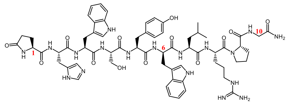
Amino acid residues at both ends of linear peptides are susceptible to degradation by exopeptidases, so modification of terminal residues by methylation or N-acetylation can protect the peptide drugs in the metabolic process.
The glucose-dependent insulinotropic polypeptide (GIP) has a half-life of 2-5 minutes. Mabilleau et al. developed an enzyme-stable GIP analog by acetylating Tyr1 (N-AcGIP) in GIP, with an in vivo half-life greater than 24h.
- Cyclization or double cyclization
The configuration of the linear peptide is unstable, and it’s easy to deform and bind to the catalytic center of protease. The cyclic method can stabilize the peptide configuration, reduce the binding constant (Ka) with protease, and lower the affinity with protease. At the same time, through cyclization, amino acid residues at both ends or metabolic sites can be protected, which becomes an effective way to enhance the stability of peptides. More than two-thirds of the peptide drugs on the market are cyclic peptide compounds. For example:
Many antibiotics of natural origin are cyclic peptides, such as cyclosporine and daptomycin. Cyclosporine is highly stable to gastrointestinal peptidases (>90% intact after 2 hours of incubation). Sandimmune® is a kind of lipid-based preparation, and Neoral® is a microemulsion with improved bioavailability. The final half-life of Neoral® ranges from 5 to 18 hours after oral administration.
Lanreotide, the 8-residue cyclic peptide analog of somatostatin, is used for the treatment of acromegaly (gigantism) and GEP-NETs caused by excessive hormone release by the pituitary gland. Researchers developed subcutaneous long-acting formulations (Somatuline Autogel® and Somatuline Depot®) that can continuously release over a month to avoid daily injections. Lanretide contains two unnatural amino acid residues at sites 1 and 4 and an amidate Thr8. The half-life is extended to 23 to 30 days after modification compared with 3 minutes for endogenous somatostatin.
PEG has a variety of drug-related properties: high water solubility and mobility, low toxicity and immunogenicity, and easy to clear from the body. Similar properties are obtained when the drug is modified with PEG, and the transfer of these properties is positively correlated with the molecular weight of PEG. PEG modification at the end of the peptide chain can not only protect the amino acid residues but also significantly increase the molecular weight and steric hindrance. Lee et al. showed that site-specific pegylation of GLP-1 can result in a 16-times increase in plasma half-life in rats.
Another important purpose of PEG modification with large molecular weight is to reduce glomerular filtration rate and decrease renal clearance. Both mechanisms work together to greatly prolong the elimination half-life in vivo.
- Add ligands to improve binding to albumin or IgG
In vivo, albumin or IgG uses the neonatal Fc receptor (FcRn) circulatory pathway to avoid degradation. When albumin is internalized into cells, it has a strong binding with FcRn at pH 6 in endosomes to protect albumin from lysosome degradation. Then, the combination is released outside the cell to a pH 7.4 environment where it releases albumin. In response to this pathway, fatty chain modification was performed in the peptide structure to bind albumin. Liraglutide and Simeglutide both significantly increased their half-lives by this method. Other research methods include covalent binding of peptide and albumin, recombinant expression, etc.
In addition to the methods above, the half-life of drugs in vivo can also be prolonged by the combination of enzyme inhibitors and the use of liposomes and other sustained-release preparations to protect free peptides.
Summary
Further understanding of the metabolic stability of peptides and early discovery of metabolic mechanisms and sites can help researchers carry out structural modifications to improve drug stability, which is very important for the success of the clinical development of peptides. Currently, many long-acting peptide drugs and slow-release delivery preparations have been successfully marketed. With the development of science and technology, including multifunctional peptides, peptide-coupled drugs, and other new molecular types, peptides will have the opportunity to be used to treat more diseases.
Related Products
| Product Name | Description |
| Triptorelin | Triptorelin is a synthetic gonadotropin-releasing hormone (GnRH) peptide agonist that binds to the GnRH receptor. It inhibits the growth of DU145, LNCaP, and PC3 prostate and OVCAR-3 ovarian cancer cells. |
| Daptomycin | Daptomycin is a cyclic lipopeptide antibiotic that inhibits GRAM-POSITIVE BACTERIA. |
References
1. Yao JF, Yang H, Zhao YZ, Xue M. Metabolism of Peptide Drugs and Strategies to Improve their Metabolic Stability. Curr Drug Metab. 2018;19(11):892-901.
2. Sharma, R.; McDonald, T.S.; Eng, H,; Limberakis, C.; Stevens, B.D.; Patel, S.; Kalgutkar, A.S. In vitro Metabolism of the Glucagon-Like Peptide-1 (GLP-1)-Derived Metabolites GLP-1(9-36)amide and GLP-1(28-36)amide in Mouse and Human Hepatocytes. Drug Metab. Dispos., 2013, 41, 2148-2157.
3. Serada M, Sakurai-Tanikawa A, Igarashi M, Mitsugi K, Takano T, Shibusawa K, Kohira T. The role of the liver and kidneys in the pharmacokinetics of subcutaneously administered teriparatide acetate in rats. Xenobiotica. 2012 Apr;42(4):398-407.
4. A. Zorzi, K. Deyle and C. Heinis, Cyclic peptide therapeutics: past, present and future, Curr. Opin. Chem. Biol., 2017, 38, 24–29.
]]>There are many types of cell-penetrating peptides (CPPs), and their classification is based on physicochemical properties, sources, intake mechanisms, and biomedical applications. There is no unified conclusion yet. According to their physical and chemical properties, CPPs can be divided into three types: cationic, amphiphilic and hydrophobic, among which cationic and amphiphilic CPPs are dominant, accounting for about 85%, while hydrophobic CPPs account for only 15%.
- Cationic Cell-Penetrating Peptides
Cationic cell-penetrating peptides (CPPs) are composed of short peptides rich in arginine, lysine and histidine, such as TAT, Penetratin, Polyarginine, P22N, DPV3 and DPV6. Among them, arginine contains a guanidine group, which is hydrogen-bonded to negatively charged phosphate groups on the cell membrane and mediates CPPs entry into the membrane at physiological PH. Studies on oligo-arginine (from 3 R to 12 R) showed that the membrane penetration capacity was increased when the amount of arginine was at least 8, and the membrane penetration capacity gradually increased with the increase of the amount of arginine. Lysine, though cationic like arginine, does not contain guanidine, so when it is alone, its transmembrane efficiency is not too high.
- Amphiphilic Cell-Penetrating Peptides
The amphiphilic cell-penetrating peptides is composed of hydrophilic and hydrophobic domains, and can be divided into primary amphiphilic, secondary α-helix amphiphilic, β-folded amphiphilic and proline-enriched amphiphilic.
- Hydrophobic Cell-Penetrating Peptides
Hydrophobic cell-penetrating peptides contain only non-polar amino acid residues, the net charge is less than 20% of the total charge of the amino acid sequence, or contain hydrophobic motifs or chemical groups that are crucial for membrane penetration. Although often overlooked, these cell-penetrating peptides do exist, such as fibroblast growth factor (K-FGF) and fibroblast growth factor 12 (F-GF12) derived from Kaposi’s sarcoma.
Applications of Cell-Penetrating Peptides
- Treatment of tumors
CPPs can use their membrane-penetrating effects to carry chemotherapeutic drugs or biologic agents into the body, and release the carrier in the cytoplasm or target action sites to maximize the effectiveness of the treatment of the resulting tumor cells. Dubikovskaya et al. linked the penetrating peptide R8 to paclitaxel (PTX), a widely used chemotherapeutic drug, through a biolysable disulfide ligand. After penetrating the cell membrane, it encountered the chemical bond of the coupling with high concentration of glutathione, resulting in its cleavage. Studies have shown that CPP-paclitaxel compounds are more effective than paclitaxel alone in both in vitro cell cultures and animal models. The activity of the drug was also observed in paclitaxel-resistant cells.
- Oligonucleotide and siRNA delivery
Peptide, protein and gene-based biomacromolecules play an important role in the treatment of many diseases. However, due to their large size and low lipophile characteristics, they cannot effectively cross the cell membrane, so the application of these biomacromolecules with therapeutic value in medical fields is greatly limited. Currently, the commonly used methods for introducing biological macromolecules into cells include liposome mediated method, virus vector mediated method, electroporation method and microinjection method, etc. However, these methods have disadvantages such as low transport efficiency, high cytotoxicity, poor safety and poor targeting specificity, which greatly limit their large-scale application. Therefore, the development of efficient and safe transmembrane transport technology of biological macromolecules has become an important challenge, and the discovery of cell-penetrating peptides (CPPs) provides a new way to solve the above problems.
Oligonucleotides and siRNA are important tools for regulating protein activation and post-transcriptional gene expression, and are important means for targeted therapy of specific genes. siRNA can play its role in gene therapy through post-transcriptional gene silencing. Li et al constructed a novel siRNA delivery system sTOLP(loaded with siRNAOA-R8 / TF-modified LNP) for cell penetrating peptioyl-octadecarginine (OA-R8) transferrin (Tf)-modified multifunctional lipid nanoparticles (LNP). siRNA encapsulated in sTOLP showed strong tumor inhibition (61.7%) and was preferentially absorbed by hepatocytes and tumor cells in HepG2-containing mice without inducing immunogenicity or hepatic or renal toxicity.
- Inflammation-targeted therapy
Nuclear transcription factor kappaB (NF-κB) is an important transcriptional regulator in cells. It usually binds to its inhibitory protein in a non-activated state in the form of P50-P65 dimer. Many inflammatory responses are strongly associated with NF-κB activation. By combining CPP (8K, octa-yLysine) with NF-κB essential regulator binding domain (NBD), Dave et al. treated mice with inflammatory bowel disease by intraperitoneal injection. The results showed that the activation of NF-κB was inhibited in the treated mice, and inflammatory cytokines TNF-α, IL-6 and IL-1 showed a decreasing trend, and intestinal inflammation was improved.
- Bactericidal effect
Broad-spectrum antibiotics are difficult to inhibit or kill intracellular bacteria, which cause great harm to animal and human health, especially zoonotic infectious diseases. Due to the selective permeability of cell membrane, drugs can not penetrate the barrier, resulting in the efficacy of the ideal state. Some researchers attempted to connect CPP to peptide nucleic acids (PNAs), which could effectively kill intracellular listeria and caenorhabditis elegans (Milletti,2012), providing ideas for the design of intracellular bactericidal drugs introduced by CPP to a certain extent. The penetrating CPPs can bind specific bactericidal active substances into cells through direct transport or endocytosis, and the specific bactericidal active substances can play a selective therapeutic role in cells, thus providing a more feasible solution for the treatment of intracellular bacteria.
- Imaging
CPPs, which uses its internalization as a carrier to deliver fluorescent particles into cells, has been a promising tool for imaging agents, contrast agents and quantum dot delivery. This imaging technique has the advantage of being able to visualize and quantify biomarkers, biochemical and intracellular processes, detect the stages of disease, identify the extent of disease and measure the effectiveness of treatment.
- Viral Delivery Peptide
CPPs can also be used to increase the transduction efficiency of viruses. CPP-modified Adv as a delivery vehicle is a promising tool for cell transduction and gene therapy.
]]>How to improve efficacy while reducing side effects? Drug conjugation technology may be a solution. Through coupling technology, on the basis of cytotoxic molecules, they are coupled with tumor-targeting components to produce a complex of multiple molecules linked together to make cytotoxic molecular targeted therapeutic drugs, which can be effective to solve the above problems.
Targeted therapy that selectively binds and exerts effects through molecules expressed on the surface of cancer cells is a major advancement in cancer treatment, because compared with traditional cytotoxic drugs, targeted therapy has higher efficacy and better tolerability.
In cancer treatment, there are three targeted methods for enhancing the specificity and anti-tumor activity of cancer treatment. Targeting agents can be designed to inhibit proteins expressed by tumor cells, such as receptors or enzymes; another method is to combine effector molecules (such as ADCs, double antibodies or CAR-T) with molecules overexpressed on the surface of tumor cells and cooperate with them. Inhibit tumor cell division, while providing cytotoxic payload or stimulating tumor-oriented immune response; the third method is to use peptide-conjugated drugs (PDC) to drive the enrichment of tumor cell toxic payload.
Antibody-drug Conjugate (ADC)
Antibody-Drug Conjugates (ADCs) are a new class of highly potent biological drugs built by attaching a small molecule anticancer drug or another therapeutic agent to an antibody, with either a permanent or a labile linker.
The first-generation ADC is represented by Pfizer’s Mylotarg, which has the disadvantages of low antigen specificity, strong toxicity, unstable linker, and short half-life. The second-generation ADC is represented by Seattle’s Adcetris and Roche’s Kadcyla. Compared with the first-generation ADC, the second-generation ADC has the advantages of strong antigen specificity, higher drug efficacy, and lower immunogenicity. However, it still has strong toxicity, drug resistance, and high DAR (drug-to- antbody ratio, antibody drug loading rate) value and other issues. The third-generation ADC is site-specifically coupled and adopts a new target to have a homogeneous and single ADC. The cytotoxic molecules are more effective, with higher accuracy and lower toxicity.
Peptide-Drug Conjugate (PDC)
Many targeted drugs currently in clinical use are based on monoclonal antibodies. However, the therapeutic application of ADC is limited by its physicochemical and pharmacodynamic properties. PDC uses peptides as tumor targeting carriers with many advantages. Compared with ADCs, they are easy to synthesize, and structural modifications can be easily introduced, supporting reasonable drug design to improve bioavailability, affinity and stability. In addition, peptides have low immunogenicity.
There are three basic elements of PDC: peptide, cytotoxin and linker. The first is the choice of peptides. In recent years, with the rapid development of technologies such as proteomics, phage display, and peptide solid-phase synthesis, more and more new peptides have been discovered and rationally designed, which has greatly promoted the development of PDC. Peptide molecules used for PDC can generally be divided into cell penetrating peptides (CPPs) and cell targeting peptides (CTPs). The former can transport drugs across the cell membrane, and the latter can specifically interact with target cells. On the receptor binding. It has been reported in the literature that CPP-drug conjugates can enter cells through transport or receptor-mediated non-endocytic transport pathways that have nothing to do with energy.
Commonly used CPPs include trans-activator of transcription (TAT), transportan, penetratin and their derivatives or other peptides that have the ability to penetrate membranes. Commonly used CTPs include arginine-glycine-aspartate (RGD) constant series peptides, luteinizing hormone releasing hormone (LHRH)-like peptides, and new tumor-targeting peptides screened by phage display technology.
The second is the choice of cytotoxic drugs. The cytotoxic drugs used for PDC coupling are usually chemotherapeutic drugs, such as paclitaxel, DOX, CTP, etc., which exert anti-tumor effects by interfering or blocking the cell proliferation process. However, the classic and commonly used chemotherapeutic drugs tend to cause damage to normal cells and tissues due to their low selectivity and poor tumor targeting ability. The formation of PDC can improve the targeting of these drugs to tumor tissues and reduce their distribution in normal tissues, thereby reducing adverse reactions.
Then last is the choice of linker. The linker is an effective bridge connecting the peptide and the drug, and the linker will affect the function of the peptide or drug. Similar to ADC, PDC linkers are divided into non-breakable and breakable types. The ideal linker should have the characteristics of low molecular weight, appropriate length, appropriate stability and polarity.
Based on the above three elements, PDC has the following advantages:
In terms of technology, PDC is a new type of conjugated drug. Its design principle is partially similar to that of ADC. It is mainly used for drug delivery and tumor targeting. The difference is that the antibody component in ADC is used as a peptide that can be used as a targeting ligand. It is precisely because PDC replaces antibodies with peptides. Compared with ADC, PDC has a smaller molecular weight. Therefore, PDC drugs have better permeability of blood vessels, tissues and cells, and are easy to penetrate deep into the tumor, and do not cause an immunogenic reaction. In addition, PDC drugs can be quickly eliminated by the kidneys, and are less toxic to bone marrow and liver; on the other hand, unlike phages, adenoviruses or other microorganisms dedicated to transporting drugs, PDC drugs do not contain infectious substances.
In terms of technology, PDC targets tumor cells with a peptide chain of about 10 amino acids, and changes the conjugated hydrophobicity and ionization by controlling the amino acid sequence of the peptide chain, both of which affect the bioavailability in vitro and in vivo. Compared with the complex process of antibody production, PDC can be prepared on a large scale by solid-phase synthesis. The synthesis and purification, storage and quality control of PDC are relatively easy, and the stability in vivo and in vitro is better, which can effectively reduce large-scale production. the cost of.
FDA approved Peptide-Drug Conjugate
In January 2018, Lutathera developed by Advanced Accelerator Applications S.A, a subsidiary of Novartis, was approved by the FDA. Lutathera is the world’s first PDC drug. Lutathera (lutetium Lu 177 dotatate) is a peptide receptor radionuclide therapy (PRRT) indicated for the treatment of somatostatin receptor positive gastroenteropancreatic neuroendocrine tumours.
In February 2021, Oncopeptides announced that Pepaxto (melphalan flufenamide, also known as melflufen) has been approved by the FDA. Melflufen is an anti-cancer PDC drug targeting aminopeptidase for the treatment of multiple myeloma. It can couple an alkylating agent with a peptide targeting aminopeptidase. Aminopeptidase is present in all human cells and is overexpressed in many tumors, including multiple myeloma.
Small molecule-drug conjugates (SMDC)
Similar to the ADC structure, there are three key factors: small molecule targeting ligands, cytotoxic molecules and linkers. Its mechanism of action is also similar to that of ADC, but SMDC can disperse into tumor tissues more quickly and evenly, with low cost and no immunogenicity. At present, SMDC still has no marketed drugs. The main difficulty in its research and development is that small molecule ligands are difficult to obtain, which restricts its development.
Radionuclide Drug Conjugates (RDC)
Radionuclide drug conjugates (RDC) are mainly composed of targeting ligands, linkers, chelates and cytotoxic/imaging factors (radioisotopes). The biggest difference between RDC and ADC and SMDC is the drug load. RDC is no longer loaded with small molecules, but radionuclides. Using different medical nuclides, they can have different functions such as imaging or treatment, and some nuclides have both functions. Based on these functions, RDC will likely become a new technology for tumor diagnosis, imaging and treatment.
]]>Peptide library is a group of a large number of several million or more peptides arranged in sequence, that is, the combination of amino acid sequences of peptides of this length. The peptide library is a series of peptides and amino acid chains, which are an important part of life. Peptide libraries can be produced by companies that specialize in peptides and other biological materials, or they can be produced in the laboratory. In a laboratory capable of synthesizing peptides, many researchers order peptide libraries from companies. Complete the order very quickly. Peptide libraries are generally available to anyone who wants to order. Like many biological materials used in research, peptides are not strictly controlled because they are not considered potentially dangerous. This also makes it easy for laboratories and researchers to quickly obtain the peptides they need. In contrast, dangerous biological materials such as viruses may be restricted.
Types of peptide libraries
The simplest combinatorial peptide library is the Parallel library. The peptides of each sequence in this peptide library are synthesized in a separate reactor to ensure the quality of the peptide library. This method has a small amount of synthesis and can improve efficiency through automation and microwave technology.
The second peptide library is the Random library, which generates millions of peptides with different sequences in each reactor, which facilitates rapid screening of a large number of peptides.
What is an overlapping peptide library
Overlapping Peptide Library-This tool allows you to generate overlapping peptides to cover the full length of the protein of interest. You can define sequence parameters, such as the length of the peptide segment and the number of amino acid offsets (see fig1). The designed sequence can be edited and exported to word or Excel format. The design of overlapping peptide library is mainly determined by the two parameters of peptide chain length and offset number. The design is gradually intercepted from the N-terminus of the target protein to the C-terminus. Each time one or a few amino acids are transferred, there is partial overlap between the sequences. Finally, an overlapping peptide library is formed.

Figure 1. What is an overlapping peptide library
Production method
By artificially synthesizing genes encoding random polypeptide segments, and then cloning these genes into phage vectors, using phage epitope display technology to display the polypeptides one by one on the surface of the phage to construct a polypeptide library. Many peptide libraries are produced on solid supports such as resins or beads, and they can also be packed in vials or test tubes. Based on the specific interactions between biological macromolecules, small molecules that can mimic the biological functions and activities of specific biological macromolecules can be screened from the library through specific immune responses similar to antigens and antibodies.
Application areas of overlapping peptide libraries
Peptide libraries can be used in a wide range of research applications, and people hope to study combinations of various amino acids. Researchers who want to identify specific peptides or understand the role of peptides in cells can also use these peptide libraries. Like those engaged in drug development, peptide libraries can be used to identify active compounds. Other uses include protein purification and the development of synthetic vaccines. The peptide library can also be used to study intracellular signaling pathways. Overlapping peptide libraries design key short peptide sequences based on the target protein or long peptide, which can be used to identify antigen peptide sequences, screen enzyme substrates, and identify T cell epitopes.
References
- Lam K S, Salmon S E, Hersh E M, et al. A new type of synthetic peptide library for identifying ligand-binding activity[J]. Nature, 1991, 354(6348): 82-84.
- Schatz P J, Cull M G, Miller J F, et al. Peptide library and screening method[J]. EP, 2000.
- Reese G, Ayuso R, Leong-Kee S M, et al. Characterization and identification of allergen epitopes: recombinant peptide libraries and synthetic, overlapping peptides[J]. Journal of Chromatography B: Biomedical Sciences and Applications, 2001, 756(1-2): 157-163.
- Li X, Wang L, Zhao D, et al. Identification of host cell binding peptide from an overlapping peptide library for inhibition of classical swine fever virus infection[J]. Virus genes, 2011, 43(1): 33-40.
Alanine scanning library is one of the peptide library design tools. Alanine Scanning Library, this tool allows you to systematically substitute alanine for each amino acid position in your peptide sequence (see Fig1). Alanine is the smallest of the naturally occurring amino acids. It can maintain its chirality. In molecular biology, alanine scanning is a site-directed mutagenesis technique used to determine the contribution of specific residues to the stability or function of a given protein. Alanine scanning library can be a powerful tool to explore the effects of single amino acid side chains on protein functions and properties. Alanine can be substituted for each non-alanine residue in turn, one at a time, and subsequently, the corresponding change in epitope activity can be measured. Substituting alanine for key amino acid residues will cause changes in epitope binding activity, so that the contribution of each amino acid to peptide function can be quickly determined.
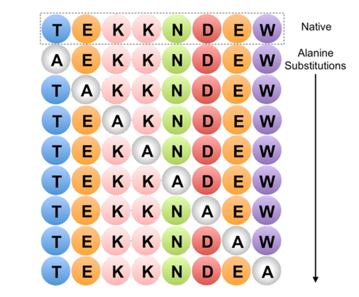
Figure 1. Example of Alanine Scanning. The native protein (top row) and each possible point mutation to alanine is considered
Fundamental
Alanine scanning mutagenesis is a powerful protein engineering technique used to study protein structure-function relationships, map binding sites, and design more stable proteins or proteins with altered properties. Alanine (Ala) scanning is a widely used mutagenesis method in which residues in the target protein are systematically substituted for alanine at selected positions by site-directed mutagenesis. Alanine is usually the accepted single-residue choice for mutation scanning because it eliminates side chain (R=methyl) interactions, but does not change the main chain conformation, nor does it impose extreme static or steric effects.
Advantages of using alanine
Alanine is used because of its small size, chemical inertness, and methyl functionality, but it mimics the secondary structure preferences of many other amino acids. Therefore, alanine scanning is particularly useful for determining the contribution of specific side chains while preserving the natural and entire protein structure. Sometimes, when the size of the mutated residue needs to be preserved, larger amino acids, such as valine or leucine, are used.
Application field
The alanine scanning library is designed to identify specific amino acid residues responsible for peptide function, stability and conformation.
A good example of alanine scanning is to examine the role of charged residues on the surface of a protein. In a systematic study on the role of conserved charged residues on the surface of the epithelial sodium channel (ENaC), alanine scanning was used to reveal the importance of charged residues in the transport of proteins to the cell surface.
Identify key epitope groups
Alanine scanning is used to simultaneously determine the functional contribution of the 19 side chains buried at the interface between the human growth hormone and the extracellular domain of its receptor. Every amino acid in the side chain is replaced by alanine. Then, a shotgun scanning method that combines the concepts of alanine scanning mutagenesis and binomial mutagenesis with phage display technology is adopted.
Assess the functionally active groups of enzymes
Another key application of alanine scanning is to determine the effect of a single residue on the structure and activity of the prototype cyclic peptide kalata B1. Cyclic peptides display a wide range of pharmaceutically important biological activities, but their natural function is to act as insecticides in plant defense. In the structure of the cyclic peptide kalata B1, all 23 non-cysteine residues are successively substituted by alanine. The data is tested by nuclear magnetic resonance spectroscopy.
References
- Morrison K L, Weiss G A. Combinatorial alanine-scanning[J]. Current opinion in chemical biology, 2001, 5(3): 302-307.
- Weiss G A, Watanabe C K, Zhong A, et al. Rapid mapping of protein functional epitopes by combinatorial alanine scanning[J]. Proceedings of the National Academy of Sciences, 2000, 97(16): 8950-8954.
- Simonsen S M, Sando L, Rosengren K J, et al. Alanine scanning mutagenesis of the prototypic cyclotide reveals a cluster of residues essential for bioactivity[J]. Journal of biological chemistry, 2008, 283(15): 9805-9813.
- Howlader M T H, Kagawa Y, Miyakawa A, et al. Alanine scanning analyses of the three major loops in domain II of Bacillus thuringiensis mosquitocidal toxin Cry4Aa[J]. Applied and environmental microbiology, 2010, 76(3): 860-865.
- Edelheit O, Hanukoglu I, Dascal N, et al. Identification of the roles of conserved charged residues in the extracellular domain of an epithelial sodium channel (ENaC) subunit by alanine mutagenesis[J]. American Journal of Physiology-Renal Physiology, 2011, 300(4): F887-F897.
Peptide microarrays (also commonly called peptide chips or peptide epitope microarrays) are designed based on protein-protein interactions, and a known protein array is immobilized on a carrier to detect paired unknown proteins. The protein immobilized on the carrier can be antibodies, antigens, receptors, ligands, enzyme substrates, and protein binding factors. This new technology can detect the relative abundance of hundreds of proteins in biological samples in a single experiment. Its core technologies include: design, modification, and high-purity synthesis of new peptide molecules that specifically bind to the target protein; high-precision, high-throughput peptide array chip design and synthesis of the target protein binding site; peptide and peptide-protein reactions detection system; antibody-specific epitope original discovery system; target protein binding peptide discovery system; and huge epitope element-binding sample library and peptide molecule sample library.
Peptide microarrays can effectively find disease-specific markers from patient biological samples and can be used for the early diagnosis of various diseases including tumors. At the same time, the use of peptide microarray technology can screen out effective substances from a large number of pharmaceutical ingredients, which is of vital significance for the design and development of new drugs and targeted individual treatment.
Determination guidelines for peptide microarrays
The determination criteria for peptide microarrays are similar to the enzyme-linked immunosorbent assay (ELISA) protocol. The peptides (thousands of several copies) are attached to the surface of the glass chip. This peptide chip can be directly cultivated with various biological samples, such as purified enzymes or antibodies, patient or animal serum, cell lysates, etc. After some washing steps, a secondary antibody with the required specificity (for example, human/mouse anti-IgG or anti-phosphotyrosine or anti-myc) is used. Typically, the secondary antibody is labeled with a fluorescent label that can be detected by a fluorescent scanner. Other detection methods include chemiluminescence or autoradiography. By analyzing the differences in the results of microarray screening of patient samples and healthy human samples, we can obtain biomarkers that are highly related to the disease, and further use these biomarkers to assist in the diagnosis, monitoring, and treatment effect evaluation of the disease.
Features of peptide microarrays
- High-throughput and systematic: a large number of different types of peptides are carried on a detection array, which can easily cover many components in the product, beyond the limitations of traditional chemical analysis methods.
- Scientificity and objectivity: Produce clear quantitative analysis results and evidence, and get rid of the subjectivity and ambiguity of empirical analysis.
- High efficiency and low cost: Thousands of peptide reagents are tested at one time, and to achieve the same results, traditional analysis methods are incomparable in terms of time and cost.
Application of peptide microarrays
- Epitope mapping: Host humoral immunological response plays an essential role in protection against pathogens. Identification of B-cell epitopes on antigens is required for accurate diagnosis and vaccine development. To map SARS-CoV-2 (severe acute respiratory syndrome coronavirus 2) spike linear epitopes, Li developed a protocol of profiling sera from patients with COVID-19 (coronavirus disease 2019) via a peptide microarray designed according to spike protein. The qualified peptide microarray exhibited bright, uniform, organized, and regular features. Using peptide microarray technology, this study provided a detailed scheme for mapping the linear epitope of SARS-CoV-2 spikes and may be applicable to linear epitope mapping of other antigens.
- Screening of epitopes: The epitope has rich biological information, which is of great value for the diagnosis of disease and the development of new vaccines. At present, the epitope research of PPRV mostly focuses on N protein and H protein. Liu used the peptide microarray technology, using the PPRV Nigeria 75/1 strain as the reference sequence, selected the full gene sequence of the four structural proteins of PPRV N, M, H, F, and combined the peptide fragments. And then they used PPRV negative-positive serum for antigen epitope screening and analyzed the response rate correlation, stability, and three modes of the obtained data.
- Rapid screening: Researchers have developed a new, simple and rapid screening method with the help of peptide microarrays to detect cell behavior in the three-dimensional tissue microenvironment. The method used electrospun fiber (polyethylene glycol) hydrogel and peptide microarray platform. The researchers studied the utility of this platform using five different peptide motifs and ten cell types. These cells included stem cells, terminally differentiated cells, cancer cells, and immune cells. These cells come from original sources or cell lines of different species. The ability of an array composed of multiple peptide motifs and concentrations to screen cell adhesion and morphology has been verified in many ways.
- Virus detection: Zhou et al. cut the extracellular sequence of 7 proteins from the HPV-16 virus in a shingled manner, constructed a microarray chip based on 241 fine peptide libraries, and performed large-scale serological screening on 75 prostate cancer patients and 80 prostate hyperplasia patients diagnosed by pathology. The significance analysis of microarrays (SAM) was used to screen the differentially expressed polypeptides (specific polypeptides) in the serum of prostate cancer patients and benign prostatic hyperplasia patients and the receiver operating characteristic curve was used to evaluate the diagnostic value of these peptides for prostate cancer and compared with PSA.
References:
- Li Y, Lai D, Tao S. SARS-CoV-2 Spike linear epitope scanning via a peptide microarray through sera profiling[J]. STAR protocols, 2021: 100707.
- Liu H. D. Screening and preliminary application of peptide epitopes of Peste des ruminants virus [D]. Gansu Agricultural University, 2018.
- Zhou Z. Application of iPDMS-based human papillomavirus type 16 (human papillomavirus, HPV-16) peptide microarray technology in the screening of serum antibodies in prostate cancer patients[D]. Soochow University, 2017.
To analyze the whole protein complement that can be expressed by a particular cell, the two dimensional- (2D) gel electrophoresis should be employed, which consist of a isoelectric focusing step in one direction of the gel and the SDS-PAGE step in the second direction of the gel. Only one sample can be analyzed on a single gel, but a higher resolution can be achieved this way. Still some protein mixtures may be too complex to be analyzed without prior fractionation; therefore most protein analysis should include some form of separation and purification before more complex analysis methods are employed. Usually any type of column chromatography, like size exclusion, ion- exchange, hydrophobic interaction or affinity chromatography is used to separate the protein mixture into smaller fractions that can then be analyzed and characterized. While the previously mentioned techniques are useful to separate and characterize a protein or peptide based on charge or molecular size, they are not capable of identifying the protein or peptide.
A traditional technique to identify the sequence of a protein or peptide is the Edman degradation procedure, where a peptide is degraded stepwise by phenylisothiocyanate, cleaved by trifluoroacetic acid and converted in aqueous HCl to phenylthiohydantoin, repeatedly until the entire protein is sequenced. Since this process is slow and has a low throughput it is no longer used in most proteomics research.
The major advancement in proteomics research came with the reliable adaptation of mass spectrometry (MS) for peptide analysis. Since most proteins are too large and complex for direct MS analysis a pre-digestion into smaller peptides by specific proteases, for example trypsin, is usually performed before analysis. In mass spectrometry the fragmented molecules are characterized by their mass to charge ratio (m/z). In order to be analyzed the particles have to be ionized into a gas-phase and are then analyzed by a mass analyzer which is coupled with a ion detector to determine the mass to charge ratio of the ion. Mass spectrometers are differentiated by their ionization source, their ion detector and mass analyzer. The most common ionization sources are MALDI (Matrix-assisted laser desorption ionization) and ESI (Electrospray ionization). Mass analyzer and ion detectors used are TOF (Time of Flight) analyzer, Quadrupol mass filter and Ion traps, such as three-dimensional Quadrupol ion trap, linear Quadrupol ion trap and orbitrap. Another common approach is the use of tandem mass spectrometry or MS/MS, where the ionized peptides are first resolved in the first mass analyzer, which separates and isolates one species at a time that is then sent to a collision cell. In the collision cell the peptide is further fragmented, and then sent to the second mass analyzer. There the mass of the fragments is precisely analyzed and a partial amino acid sequence of the peptide can be determined. These partial sequences, called peptide sequence tags, can then be used in database searches to identify the protein or peptide.
The final step after mass spectrometry analysis is to identify the protein or peptide sequence by database search or De novo sequencing. The large amount of peptide sequence tags, generated by the mass spectrometer can be compared to a database of known protein and peptide sequences to identify the actual proteins and peptide present in the sample. Databases like UniProt or SwissProt contain large amounts of information regarding protein and peptide sequences for a large variety of species. Search engines like SEQUEST or PEAKS are employed to compare the mass spectrometry results to the databases and identify the proteins with a certain amount of accuracy, defined by probability parameters. To identify the error rate of false identification of certain amino acid sequences a decoy database search can be employed, where, the mass spectra are compared to the original database and a database where the amino acid sequences are reversed. A match in both databases would be considered false and increase the False Discovery Rate (FDR) of the protein or peptide detection.
When the protein or peptide sequence is not known in any database De novo sequencing can be employed to determine the peptide amino acid sequence. De novo means “anew” or “all over” and describes the fact that the peptide has to be characterized without additional knowledge. In this regard the Edman degradation can be characterized as De novo sequencing. In mass spectrometry a labeling technique for the C-terminal amino acid can be used to identify the side by the additional mass in the mass spectrometer and the correct sequence of a peptide can be analyzed by tandem mass spectrometry. Of course the more information is available of the original protein or peptide the more accurate are the results of the mass spectrometry analysis.
Reference:
Crynen, S. (2011). Bioactive properties of peptides derived from enzymatic hydrolysis of cod muscle myosin with trypsin, chymotrypsin and elastase. University of Florida.
]]>The peptide deformylase (PDF) is a unique feature of prokaryotes and believed not to exist in eukaryotes up to even only a few years ago. PDF is essential for ribosomal protein translation by deformylating the initiating N-formylmethionine in prokaryotes. Since the initiating methionine of eukaryotes is not N-formylated, PDF inhibitors were considered as a possible antibacterial drug target. Many researchers have studied PDF and PDF inhibitors to overcome the drug resistance problem. Currently, there are 280 papers on the structure and function of bacterial PDF, and 103 papers on bacterial PDF inhibitors. Among them, LBM415 and BB-83698 are already under clinical study as novel antibiotics candidates.
However, PDF also exists in the mitochondria of eukaryotes including humans, and is crucial for cell growth according to recent papers. The first eukaryotic PDF was reported to be in the mitochondria and plastids of Arabidopsis thaliana. A bioinformatics study found many PDF-like genes in parasites, plants, and mammals. In 2004, Lee and coworkers reported that non-peptidyl bacterial PDF inhibitor actinonin suppresses the proliferation of cancer cells more than normal cells through a tumor-specific mitochondrial membrane depolarization and ATP depletion mechanism by interacting with HsPDF. It also could remove the N-formyl capping of model human mitochondrial peptides in prokaryotes. However, the function of HsPDF in vivo was unclear. The crystal structure of HsPDF was determined by EscobarAlvarez et al. and the mechanism of action was proposed. It is known that HsPDF processes formylated peptides derived from mitochondrial DNA-encoded proteins by a Co2+ dependent manner. The bacterial PDF and HsPDF have both similarities and differences in their structures. The complex structure of HsPDF and actinonin, a naturally occurring inhibitor derived from the Streptomyces species, shows an overall structural similarity to the complex structure of bacterial PDF and actinonin, but they have structural differences at the C-terminus.
In the previous study, we made novel bacterial PDF inhibitors, PMT analogs, which named after ProMediTech Ltd. In the progress of developing PMT analogs, we discovered about 600 kinds of compounds in our library. PMT analogs have potent antibacterial activity against Streptococcus pneumoniae, Haemophilus influenzae, and Moraxella catarrhalis. These bacteria were known to cause respiratory tractassociated infections. Structural modification of PMT analogs makes these compounds more effective especially against Staphylococcus aureus. PMT analogs have a peptidomimetic structure like actinonin, but have stronger activity and better pharmacokinetic properties than actinonin.
Based on our previous studies, we predicted that PMT analogs could bind to HsPDF and inhibit the proliferation of cancer cells like actinonin. In order to prove our hypothesis, we carried out virtual screening of PMT analogs with GOLD software and DOCK6.5 from UCSF and selected 43 compounds from our library. The MTT assay was performed to test the antitumor activity for 10 highly scored compounds out of the 43 compounds. Among the 10 compounds, 4 analogs, PMT-172, PMT-173, PMT-199, and PMT-201, showed similar or better activity compared to actinonin. We prepared a HsPDF construct that has an enhanced expression level by codon optimization in E. coli. Then, 1H-CPMG-T2 filter NMR experiments were performed with purified protein, and it was confirmed that PMT-172 and PMT-199 could bind directly to HsPDF. The docking models between PMT-172, PMT-199 and HsPDF from the X-ray crystal structure (PDB 3G5K) with DOCK6.5 were obtained. It was identified that PMT-172 and PMT-199 have an N-alkyl amide and sulfonyl amide group, respectively, at the functional group R3 and R4. Understanding the structural characteristics of PMT-172 and PMT-199 might yield clues to design novel anticancer drug candidates that have better physical properties and binding affinities through modification of the functional groups.
Reference:
Kim, Won-Je, et al. “Bacterial peptide deformylase inhibitor PMT analogs inhibit cancer cell growth by interacting with human peptide deformylase.” Chinese science bulletin 59.32 (2014): 4274-4282.
]]>