In recent years, more and more studies have described the antioxidant capacity of plant protein-derived peptides. Plant proteins are considered a new source of antioxidant peptides that can not only save energy, but also enhance the treatment of oxidation-related diseases, delay the oxidation of food, and thus improve the quality of life. Despite an increasing number of reports on the preparation, structure, and activity of antioxidant peptides, and the commercialization of some antioxidant peptides, a systematic review and evaluation of plant protein-derived antioxidant peptides is still lacking. This article summarizes the latest advances in plant protein-derived antioxidant peptides, including (1) the preparation, purification, identification, and application of plant protein-derived antioxidant peptides by enzymatic hydrolysis; (2) Influencing factors, evaluation methods, and mechanism of antioxidant protein hydrolysates or peptides.
Traditional purification and identification methods of antioxidant peptides are expensive and time-consuming. To solve this problem, researchers have conducted extensive exploration and research on computer-aided mathematical models (molecular docking, QSAR studies, etc.), bioinformatics tools, and real-time updated protein databases. In addition, the authors of the reviews believe that future research should focus on the large-scale commercial production of plant protein-derived antioxidant peptides, comprehensive studies on the structure-activity relationship of antioxidant peptides, an in-depth understanding of antioxidant mechanisms, and confirmation of their health benefits in vivo.
What is the plant protein-derived antioxidant peptide?
Antioxidant peptides are specific protein fragments possessing antioxidant activity and antioxidant peptides derived from plant protein are called plant protein-derived antioxidant peptides. With increasing consumer demand for high nutritional and functional proteins, researchers are shifting their focus from traditional raw proteins to plant-based proteins. In particular, natural antioxidant peptides derived from plant proteins have attracted worldwide attention due to their advantages of environmental protection, sustainability, low cost, and non-toxic side effects.
Evaluation methods and mechanism of antioxidant peptides
The effective prevention of oxidative stress and food oxidation of antioxidant peptides attracted researchers to evaluate their antioxidant capacity. At present, there are three methods to evaluate the antioxidant capacity of peptides: chemical method, cell biology method in vitro, and animal experiment in vivo. The potential mechanisms of antioxidant peptides are as follows: (1) direct scavenging of reactive oxygen species; (2) chelated metal ions; (3) Inhibition of lipid peroxidation; (4) Activate the body’s antioxidant defense system.
Enzymatic preparation of plant protein-derived antioxidant peptides
From the point of view of sustainable development and utilization of biological resources, protein resources in the by-products or wastes produced during plant processing have great potential to produce bioactive peptides. The rational development and utilization of plant processing by-products or wastes are beneficial to improve the nutritional utilization value of wastes, save energy and protect the environment. In particular, wheat gluten, wheat germ, rapeseed, watermelon seed, corn gluten, rice bran, oat bran, palm kernel cake, and wine-making grains have been used to produce antioxidant peptides.
Plant proteases (animal proteases, plant proteases, and microbial proteases) are hydrolyzed first to form proteolysis, followed by further purification, membrane separation, electrophoresis, chromatography (gel permeation chromatography (GPC), ion exchange chromatography (IEC), reversed-phase high-performance liquid chromatography (RP-HPLC), etc.) to obtain antioxidant peptides. The obtained antioxidant peptides were identified by mass spectrometry or mass spectrometry/mass spectrometry, and their antioxidant activities were evaluated by in vitro and in vivo experiments. At present, the main preparation methods for peptides are chemical extraction, fermentation, synthesis, and enzymolysis.
Purification and identification of plant protein-derived antioxidant peptide
The enzymolysis product of proteins is a mixture consisting of partially unhydrolyzed proteins, peptides with different chain lengths, hydrophobicity and net charge, and free amino acids. To evaluate the structure and activity of antioxidant peptides more accurately, it is necessary to develop suitable purification methods. At present, the main separation and purification technologies of peptides include chromatography (GPC, IEC, RP-HPLC), membrane separation, and a combination of these technologies. Identification of protein hydrolysates is usually accomplished by mass spectrometry, mass spectrometry/mass spectrometry, and bioinformatics techniques.
After a series of isolation and purification schemes, the peptide structure needs to be identified. DNA translation, mass spectrometry, and peptide sequencing are commonly used to identify peptides. It is worth noting that mass spectrometry has been widely used in the identification of peptide sequences because of its high efficiency, sensitivity, and good reproducibility. In addition, several online databases have become popular for researching antioxidant peptides. Table 2 summarizes the main bioinformatics analysis tools.
Factors influencing the action of antioxidant peptides
It is important to summarize the properties and activities of plant-derived antioxidant peptides as comprehensively as possible for guiding and predicting the relationship between the structure and function of plant-derived antioxidant peptides. The activity of antioxidant peptides is mainly affected by amino acid composition, sequence, and molecular weight.
Analysis of the structure-activity relationship of antioxidant peptides by computer-aided molecular modeling
QSAR is a mathematical model to establish quantitative dependence between the structure-activity relationships of a series of compounds. QSAR is used to evaluate the correlation between the molecular structure and activity of antioxidant peptides, which provides a good theoretical basis for the prediction, isolation, and purification of peptide activity and elucidation of molecular mechanism.
Future research direction and existing problems
As mentioned earlier, proteins derived from plant products can be used to produce antioxidant peptides. On this basis, the research progress of plant-derived antioxidant peptides was reviewed in this paper. The molecular mechanism of these peptides at the level of the whole gene will be verified with the advancement of science and technology in the future and the continuous investment in the research of these peptides in vivo. In addition, the preparation of nanoparticles and microcapsules of antioxidant peptides and their synergies with other active ingredients are also the focus of future research. Future research and industrial cooperation will help achieve the long-term goal of commercializing plant antioxidant peptides.
]]>Summary of peptides for gastrointestinal studies
1. Motilin
Motilin is a 22-amino-acid hormone, released by the enteroendocrine M cells of the duodenum and proximal jejunum.
| Name | CAS | Sequence |
| Motilin, canine | 85490-53-5 | FVPIFTHSELQKIREKERNKGQ |
| Motilin (26-47), human, porcine | 52906-92-0 | FVPIFTYGELQRMQEKERNKGQ |
2. Gastrin
Gastrin is produced by G cells located in the gastric antrum and is the primary endocrine regulator of the secretory phase of a protein meal. The main function of gastrin is to stimulate gastric acid secretion, promote gastrointestinal motility, and participate in the maintenance of iron homeostasis. At the same time, gastrin can also stimulate the secretion of the pancreas, bile, and intestinal juice, and further decompose the food in the small intestine, which is beneficial to the absorption of nutrients in the small intestine.
| Name | CAS | Sequence |
| Gastrin I (human) | 10047-33-3 | Pyr–GPWLEEEEEAYGWMDF-NH2 |
| [Leu15]-Gastrin I (human) | 39024-57-2 | Pyr-GPWLEEEEEAYGWLDF |
| Gastrin I (1-14), human | 100940-57-6 | Glp-GPWLEEEEEAYGW |
| CCK-4 Acetate | 35144-91-3 | Trp-Met-Asp-Phe-NH2 |
| Pentagastrin | 5534-95-2 | Boc-β-Ala-Trp-Met-Asp-Phe-NH2 |
| Mini Gastrin I, human | 54405-27-5 | LEEEEEAYGWMDF-NH2 |
| Big Gastrin-1, human | 60675-77-6 | Pyr-LGPQGPPXLVADPSKKQGPWLEEEEEEAYGWMDF-NH2 |
| Gastrin I rat | 81123-06-0 | XRPPMEEEEEAYGWMDF |
3. Bombesin
Bombesin (BBS) is a 14-amino-acid peptide first isolated from the skin of the frog, Bombina bombina, in 1970 by Erspamer. It stimulates the release of gastrin and pancreatic enzymes and causes contraction of the gallbladder.
| Name | CAS | Sequence |
| Bombesin | 31362-50-2 | Glp-QRLGNQWAVGHLM-NH2 |
| Bombesin nonapeptide | 55750-00-0 | NQWAVGHLM |
4. Tachykinin
The tachykinins are a family of small peptides sharing a common C-terminal sequence, Phe-Xaa-Gly-Leu-Met-NH2, whose main members are SP, neurokinin A, and neurokinin B.
| Name | CAS | Sequence |
| Eledoisin | 69-25-0 | XPSKDAFIGLM |
| Kassinin | 63968-82-1 | DVPKSDQFVGLM |
| Neurokinin A | 86933-74-6 | HKTDSFVGLM |
| Neurokinin A(4-10) | 97559-35-8 | DSFVGLM-NH2 |
| Neurokinin B | 86933-75-7 | DMHDFFVGLM-NH2 |
| Substance P (1-7) | 68060-49-1 | RPKPQQF |
5. Somatostatin
Somatostatin belongs to the expanding family of small regulatory peptides which exert a variety of actions in different organ systems throughout the body.
| Name | CAS | Sequence |
| Octreotide | 83150-76-9 | FCFWKTCT (Disulfide bridge: Cys2-Cys7) |
| Octreotide Acetate | 79517-01-4 | FCFWKTCT |
6. Vasoactive intestinal peptide (VIP) and related peptides
The vasoactive intestinal polypeptide (VIP) is a member of a family of homologous neuro- and endocrine peptides which has three separate groups distinguished by the receptors which recognize them.
| Name | CAS | Sequence |
| Prepro VIP (111-122), human | 123025-94-5 | VSSNISEDPVPV |
| Prepro VIP (81-122), human | 111366-38-2 | HADGVFTSDFSKLLGQLSAKKYLESLMGKRVSSNISEDPVPV |
| VIP(6-28)(human, rat, porcine, bovine) | 69698-54-0 | FTDNYTRLRKQMAVKKYLNSILN-NH2 |
| [D-p-Cl-Phe6,Leu17]-VIP | 102805-45-8 | HSDAVXXDNYXRLRKQLAVKKYLNSXLN |
7. Neurotensin
Neurotensin is a 13–amino acid peptide originally detected in the bovine hypothalamus. Neurotensin can protect the airway from gastric mucosal injury caused by cold stimulation and can inhibit gastric acid secretion.
| Name | CAS | Sequence |
| Neurotensin(8-13) | 60482-95-3 | RRPYIL |
| Neurotensin | 39379-15-2 | Pyr-LYENKPRRPYIL |
| [Gln4]-Neurotensin | 61445-54-3 | Pyr-LYQNKPRRPYIL |
| Kinetensin | 103131-69-7 | IARRHPYFL |
| Xenopsin | 51827-01-1 | Glp-GKRPWIL |
8. Cholecystokinin
Cholecystokinin is secreted by cells of the upper small intestine. Its secretion is stimulated by the introduction of hydrochloric acid, amino acids, or fatty acids into the stomach or duodenum.
| Name | CAS | Sequence |
| Cholecystokinin Octapeptide (desulfated) | 25679-24-7 | DYMGWMDP |
| CCK-4 Acetate | 35144-91-3 | Trp-Met-Asp-Phe-NH2 |
9. Secretin
Secretin is a gastrointestinal peptide and a member of the pituitary adenylate cyclase-activating polypeptide (PACAP)/glucagon superfamily Sherwood et al.
| Name | CAS | Sequence |
| Secretin (28-54), human | 108153-74-8 | HSDGTFTSELSRLREGARLQRLLQGLV-NH2 |
| Secretin Acetate | 10813-74-8 | HSDGTFTSELSRLRDSARLQRLLQGLV-NH2 |
| Secretin (33-59), rat | 121028-49-7 | HSDGTFTSELSRLQDSARLQRLLQGLV-NH2 |
| Secretin, canine | 110786-77-1 | HSDGTFTSELSRLRESARLQRLLQGLV-NH2 |
| Secretin (5-27), porcine | 19665-15-7 | TFTSELSRLRDSARLQRLLQGLV-NH2 |
10. Pancreatic polypeptide
Pancreatic polypeptide (PP) is a 36–amino acid, 4.2-kDa polypeptide secreted by the islet F cells. PP belongs to the peptide YY/neuropeptide Y family of polypeptides.
| Name | CAS | Sequence |
| Pancreatic Polypeptide, bovine | 179986-89-1 | APLEPEYPGDNATPEQMAQYAAELRRYINMLTRPRY-NH2 |
| Pancreatic Polypeptide, human | 75976-10-2 | APLEPVYPGDNATPEQMAQYAADLRRYINMLTRPRY-NH2 |
| Pancreatic Polypeptide, rat | 90419-12-8 | APLEPMYPGDYATHEQRAQYETQLRRYINTLTRPRY-NH2 |
11. Peptide YY
Peptide YY (PYY) is a 36-amino-acid gut hormone released by L cells of the ileum/colon in proportion to ingested calories. PYY is functionally related to NPY and binds to the Y family of receptors, Y1R–Y6R.
| Name | CAS | Sequence |
| Peptide YY (3-36) | 126339-09-1 | IKPEAPGEDASPEELNRYYASLRHYLNLVTRQRY-NH2 |
| Peptide YY (3-36) Human | 123583-37-9 | IKPEAPGEDASPEELNRYYASLRHYLNLVTRQRY |
| Peptide YY (human) | 118997-30-1 | YPIKPEAPGEDASPEELNRYYASLRHYLNLVTRQRY-NH2 |
12. Neuropeptide Y
Neuropeptide Y (NPY) is a vasoconstricting peptide that is released together with NE from sympathetic nerve endings.
| Name | CAS | Sequence |
| Neuropeptide Y (human, rat) | 90880-35-6 | YPSKPDNPGEDAPAEDMARYYSALRHYINLITRQRY-NH2 |
| Neuropeptide Y (13-36), amide, human | 122341-40-6 | PAEDMARYYSALRHYINLITRQRY-NH2 |
| Neuropeptide Y (22-36) | 119019-65-7 | SALRHYINLITRQRY-NH2 |
| Neuropeptide Y(29-64) | 303052-45-1 | YPSKPDNPGEDAPAEDMARYYSALRHYINLITRQRY |
| Neuropeptide Y, free acid, human, rat | 99575-89-0 | YPSKPDNPGEDAPAEDMARYYSALRHYINLITRQRY |
13. Opioid peptide
Opioid peptides are the endogenous ligands at opioid receptors.
| Name | CAS | Sequence |
| [D-Ala2]-Leu-Enkephalin-Arg | 81733-79-1 | Tyr-D-Ala-Gly-Phe-Leu-Arg |
| Dynorphin A(1-13) | 72957-38-1 | YGGFLRRIRPKLK |
| Dynorphin B (1-13) | 83335-41-5 | YGGFLRRQFKVVT |
| Met-Enkephalin | 58569-55-4 | YGGFM |
| N-Acetyl-α-Endorphin | 88264-63-5 | Ac-YGGFMTSEKSQTPLVT |
| β-Endorphin rat | 77367-63-6 | YGGFMTSEKSQTPLVTLFKNAIIKNVHKKGQ |
| β-Casomorphin, human | 102029-74-3 | YPFVEPI |
| β-Casomorphin (1-3), amide | 80705-23-3 | YPF-NH2 |
| β-Casomorphin (1-5), amide, bovine | 83936-23-6 | Y-d-Ala-FPM-NH2 |
| β-Casomorphin (1-6), bovine | 77434-43-6 | YPFPGP |
| β-Casomorphin (1-7), bovine | 72122-62-4 | YPFPGPI |
| α-Neoendorphin 1-8 | 83339-89-3 | YGGFLRKY |
| β-Neoendorphin | 77739-21-0 | YGGFLRKYP |
14. Gastric Inhibitory Polypeptide
GIP is a 42-amino-acid peptide hormone encoded by a single gene. GIP mRNA is expressed only in the gut, specifically in the K cells, the majority of which are located in the proximal duodenum.
| Name | CAS | Sequence |
| GIP (human) | 100040-31-1 | YAEGTFISDYSIAMDKIHQQDFVNWLLAQKGKKNDWKHNITQ |
| GIP (1-39) | 725474-97-5 | YAEGXFXSDYSXAMDKXRQQDFVNWLLAQKGKKSDWKHN |
- Replace L-type amino acids with D-type amino acids or unnatural amino acids
The main recognition site of protease in vivo is natural L-type amino acid, so replacing L-type amino acid in the metabolic site with D-type amino acid can weaken the affinity with protease and maintain the physiological activity of the peptide.
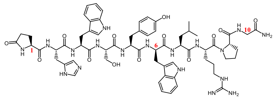
Triptorelin is a gonadotropin-releasing hormone (GnRH) analog. It replaces positions 1, 6, and 10 of the natural GnRH with three unnatural amino acid residues. Results suggest that its half-life after intravenous administration is extended to 2.8 hours compared with 5 minutes of the natural GnRH. And it’s mainly cleared by the liver and kidney.
Amino acid residues at both ends of linear peptides are susceptible to degradation by exopeptidases, so modification of terminal residues by methylation or N-acetylation can protect the peptide drugs in the metabolic process.
The glucose-dependent insulinotropic polypeptide (GIP) has a half-life of 2-5 minutes. Mabilleau et al. developed an enzyme-stable GIP analog by acetylating Tyr1 (N-AcGIP) in GIP, with an in vivo half-life greater than 24h.
- Cyclization or double cyclization
The configuration of the linear peptide is unstable, and it’s easy to deform and bind to the catalytic center of protease. The cyclic method can stabilize the peptide configuration, reduce the binding constant (Ka) with protease, and lower the affinity with protease. At the same time, through cyclization, amino acid residues at both ends or metabolic sites can be protected, which becomes an effective way to enhance the stability of peptides. More than two-thirds of the peptide drugs on the market are cyclic peptide compounds. For example:
Many antibiotics of natural origin are cyclic peptides, such as cyclosporine and daptomycin. Cyclosporine is highly stable to gastrointestinal peptidases (>90% intact after 2 hours of incubation). Sandimmune® is a kind of lipid-based preparation, and Neoral® is a microemulsion with improved bioavailability. The final half-life of Neoral® ranges from 5 to 18 hours after oral administration.
Lanreotide, the 8-residue cyclic peptide analog of somatostatin, is used for the treatment of acromegaly (gigantism) and GEP-NETs caused by excessive hormone release by the pituitary gland. Researchers developed subcutaneous long-acting formulations (Somatuline Autogel® and Somatuline Depot®) that can continuously release over a month to avoid daily injections. Lanretide contains two unnatural amino acid residues at sites 1 and 4 and an amidate Thr8. The half-life is extended to 23 to 30 days after modification compared with 3 minutes for endogenous somatostatin.
PEG has a variety of drug-related properties: high water solubility and mobility, low toxicity and immunogenicity, and easy to clear from the body. Similar properties are obtained when the drug is modified with PEG, and the transfer of these properties is positively correlated with the molecular weight of PEG. PEG modification at the end of the peptide chain can not only protect the amino acid residues but also significantly increase the molecular weight and steric hindrance. Lee et al. showed that site-specific pegylation of GLP-1 can result in a 16-times increase in plasma half-life in rats.
Another important purpose of PEG modification with large molecular weight is to reduce glomerular filtration rate and decrease renal clearance. Both mechanisms work together to greatly prolong the elimination half-life in vivo.
- Add ligands to improve binding to albumin or IgG
In vivo, albumin or IgG uses the neonatal Fc receptor (FcRn) circulatory pathway to avoid degradation. When albumin is internalized into cells, it has a strong binding with FcRn at pH 6 in endosomes to protect albumin from lysosome degradation. Then, the combination is released outside the cell to a pH 7.4 environment where it releases albumin. In response to this pathway, fatty chain modification was performed in the peptide structure to bind albumin. Liraglutide and Simeglutide both significantly increased their half-lives by this method. Other research methods include covalent binding of peptide and albumin, recombinant expression, etc.
In addition to the methods above, the half-life of drugs in vivo can also be prolonged by the combination of enzyme inhibitors and the use of liposomes and other sustained-release preparations to protect free peptides.
Summary
Further understanding of the metabolic stability of peptides and early discovery of metabolic mechanisms and sites can help researchers carry out structural modifications to improve drug stability, which is very important for the success of the clinical development of peptides. Currently, many long-acting peptide drugs and slow-release delivery preparations have been successfully marketed. With the development of science and technology, including multifunctional peptides, peptide-coupled drugs, and other new molecular types, peptides will have the opportunity to be used to treat more diseases.
Related Products
| Product Name | Description |
| Triptorelin | Triptorelin is a synthetic gonadotropin-releasing hormone (GnRH) peptide agonist that binds to the GnRH receptor. It inhibits the growth of DU145, LNCaP, and PC3 prostate and OVCAR-3 ovarian cancer cells. |
| Daptomycin | Daptomycin is a cyclic lipopeptide antibiotic that inhibits GRAM-POSITIVE BACTERIA. |
References
1. Yao JF, Yang H, Zhao YZ, Xue M. Metabolism of Peptide Drugs and Strategies to Improve their Metabolic Stability. Curr Drug Metab. 2018;19(11):892-901.
2. Sharma, R.; McDonald, T.S.; Eng, H,; Limberakis, C.; Stevens, B.D.; Patel, S.; Kalgutkar, A.S. In vitro Metabolism of the Glucagon-Like Peptide-1 (GLP-1)-Derived Metabolites GLP-1(9-36)amide and GLP-1(28-36)amide in Mouse and Human Hepatocytes. Drug Metab. Dispos., 2013, 41, 2148-2157.
3. Serada M, Sakurai-Tanikawa A, Igarashi M, Mitsugi K, Takano T, Shibusawa K, Kohira T. The role of the liver and kidneys in the pharmacokinetics of subcutaneously administered teriparatide acetate in rats. Xenobiotica. 2012 Apr;42(4):398-407.
4. A. Zorzi, K. Deyle and C. Heinis, Cyclic peptide therapeutics: past, present and future, Curr. Opin. Chem. Biol., 2017, 38, 24–29.
]]>Sources of Antimicrobial Peptides
- Microorganisms-Derived Antimicrobial Peptides
Microorganisms-derived antimicrobial peptides are classified into two types: antimicrobial peptides derived from bacterial and antimicrobial peptides derived from viral. Among them, antibacterial peptides derived from bacteria are also called bacteriocins. Bacteriocins are a type of peptide or precursor peptide molecules with bactericidal biological activity that are produced by bacteria during the metabolic process through protein. Usually, these peptides are all cationic amino acid chains, which are hydrophobic or amphiphilic. According to biochemical properties, bacteriocins can be divided into three categories: class I bacteriocins, class II bacteriocins and class III bacteriocins. Among them, type I bacteriocins are also called thioether antibiotics, with a molecular mass of less than 5ku and a number of amino acids of about 19 to 38. They have mucosal activity and good heat resistance. Their structure contains rare thioether amino acids, such as Lanthionine and β-methyllanthione. Class II bacteriocins are a type of antibacterial peptides called lactic acid bacteriocins discovered in recent years. Based on their biological, physical and chemical properties, they can be used as food preservation agents. Compared with class I bacteriocins, class II bacteriocins have a slightly smaller molecular mass (4-6 ku). Class III bacteriocins are secreted by bacterial proprotein translocases, with a molecular weight greater than 30 ku and good heat resistance. The other type is antibacterial peptides derived from viral, which are currently found in relatively few types. Among them, LLPs are lentiviral cytolytic peptides encoded by the C-terminal sequence of the human defect virus type 1 (HIV-1) transmembrane protein, which have strong toxic effects on microorganisms and cells. LLPs have high arginine and do not contain lysine. In most cases, microorganisms-derived antimicrobial peptides exist in a cyclic structure.
- Plant-Derived Antimicrobial Peptides
Plant-derived antibacterial peptides have active effects on plant pathogens and bacterial pathogens that infect humans. They are a barrier to the plant defense system and considered to be antibiotic peptides with important application. Common plant-derived antibacterial peptides include thionin, defensins, lipid transfer proteins, cell penetrating peptides, ecdysone, etc. They have similar characteristics, such as positive charge, disulfide bond, and the target sites are all located in the outer cell membrane and so on.
- Insect-Derived Antimicrobial Peptides
Insects are the most abundant creatures in the biological world, with more than 1 million species. They also have strong adaptability and developed defense mechanisms. Antimicrobial peptides are a type of peptide produced by the hemolymph or related parts of the body when insects are stimulated by the external environment. They are an organic part of the insect’s humoral immune system and play an important role in the entire immune system. Antibacterial peptides secreted by its fat body inhibit pathogens in various systems. So far, there are about 200 kinds of antibacterial peptides isolated in insects. These peptides are divided into 5 types according to their antibacterial mechanism and amino acid sequence: cecropins, lysozyme, defensins, glycine-rich peptides and proline-rich peptides.
- Amphibian-Derived Antimicrobial Peptides
The skin of amphibians is exposed, the humidity is high, and it has respiratory function. This morphological and physiological characteristic requires that it can only survive in a humid environment, which is also an excellent environment for some pathogenic microorganisms to survive. In order to exploit and adapt to vast habitats and diverse ecological environments, amphibians eventually formed a defense system against pathogenic microorganisms through continuous evolution. Antibacterial peptides, an important effector molecule of innate immunity released by skin glands, play an important role in the immune defense mechanism of amphibians when they are stimulated by adrenaline, stress, trauma and other factors. After long-term evolution, the antimicrobial peptide gene has undergone multiple duplications and mutations, resulting in a large number of structures and a wide variety of antimicrobial peptides. At present, 1,400 antimicrobial peptides have been isolated from amphibian skin secretions, and secretions from different sources contain different antimicrobial bioactive molecules. Amphibian-derived antimicrobial peptides can be divided into two categories according to their structural characteristics: cyclic antimicrobial peptides containing intramolecular disulfide bonds and linear antimicrobial peptides with α-helical structure.
- Mammalian Antimicrobial Peptides
Antimicrobial peptides in mammals only have a lethal effect on exogenous bacteria, but are harmless to normal cells of the animal. Mammalian antimicrobial peptides mainly exist in mucosal epithelial cells and neutrophils and skin, such as surfactant anionic antimicrobial peptides in the respiratory tract, and defensins in epithelial tissues. These antimicrobial peptides and phagocytes jointly establish the body’s first line of defense. Mammalian antimicrobial peptides are mainly divided into two categories, namely defensins and cathelicidins. The most studied defensin antimicrobial peptides are the largest class of antimicrobial peptides. Mammalian defensins are divided into two categories: α-defensins and β-defensins.
- Marine Antimicrobial Peptides
The ocean is the origin of life and a huge living place for creatures. It is estimated that about 1/2 of the world’s creatures live in the ocean. This huge biobank contains abundant active antibacterial substances. It has been proved before that a wide variety of marine organisms can produce a variety of antibacterial peptides, of which the research on marine animal antibacterial peptides accounts for the majority. Among marine vertebrates, fishes are the most reported to secrete antimicrobial peptides. Due to the specificity of its body structure, antimicrobial peptides are mainly secreted into the mucus layer of marine fish, which act as the first barrier of the non-specific immune system to kill pathogenic bacteria before entering the organism. Marine biological antimicrobial peptides have the characteristics of rapid synthesis and secretion, fast diffusion and flexibility, which are beyond the functions of other immune system. Marine invertebrates such as molluscs, tunicates, crustaceans, etc. mainly rely on the innate immune mechanism of humoral immunity and cellular responses as defense systems. Among them, antimicrobial peptides, as an important component of the innate immune defense system, can respond quickly and in real time to invading pathogenic microorganisms.
]]>Alanine scanning library is one of the peptide library design tools. Alanine Scanning Library, this tool allows you to systematically substitute alanine for each amino acid position in your peptide sequence (see Fig1). Alanine is the smallest of the naturally occurring amino acids. It can maintain its chirality. In molecular biology, alanine scanning is a site-directed mutagenesis technique used to determine the contribution of specific residues to the stability or function of a given protein. Alanine scanning library can be a powerful tool to explore the effects of single amino acid side chains on protein functions and properties. Alanine can be substituted for each non-alanine residue in turn, one at a time, and subsequently, the corresponding change in epitope activity can be measured. Substituting alanine for key amino acid residues will cause changes in epitope binding activity, so that the contribution of each amino acid to peptide function can be quickly determined.
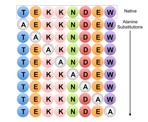
Figure 1. Example of Alanine Scanning. The native protein (top row) and each possible point mutation to alanine is considered
Fundamental
Alanine scanning mutagenesis is a powerful protein engineering technique used to study protein structure-function relationships, map binding sites, and design more stable proteins or proteins with altered properties. Alanine (Ala) scanning is a widely used mutagenesis method in which residues in the target protein are systematically substituted for alanine at selected positions by site-directed mutagenesis. Alanine is usually the accepted single-residue choice for mutation scanning because it eliminates side chain (R=methyl) interactions, but does not change the main chain conformation, nor does it impose extreme static or steric effects.
Advantages of using alanine
Alanine is used because of its small size, chemical inertness, and methyl functionality, but it mimics the secondary structure preferences of many other amino acids. Therefore, alanine scanning is particularly useful for determining the contribution of specific side chains while preserving the natural and entire protein structure. Sometimes, when the size of the mutated residue needs to be preserved, larger amino acids, such as valine or leucine, are used.
Application field
The alanine scanning library is designed to identify specific amino acid residues responsible for peptide function, stability and conformation.
A good example of alanine scanning is to examine the role of charged residues on the surface of a protein. In a systematic study on the role of conserved charged residues on the surface of the epithelial sodium channel (ENaC), alanine scanning was used to reveal the importance of charged residues in the transport of proteins to the cell surface.
Identify key epitope groups
Alanine scanning is used to simultaneously determine the functional contribution of the 19 side chains buried at the interface between the human growth hormone and the extracellular domain of its receptor. Every amino acid in the side chain is replaced by alanine. Then, a shotgun scanning method that combines the concepts of alanine scanning mutagenesis and binomial mutagenesis with phage display technology is adopted.
Assess the functionally active groups of enzymes
Another key application of alanine scanning is to determine the effect of a single residue on the structure and activity of the prototype cyclic peptide kalata B1. Cyclic peptides display a wide range of pharmaceutically important biological activities, but their natural function is to act as insecticides in plant defense. In the structure of the cyclic peptide kalata B1, all 23 non-cysteine residues are successively substituted by alanine. The data is tested by nuclear magnetic resonance spectroscopy.
References
- Morrison K L, Weiss G A. Combinatorial alanine-scanning[J]. Current opinion in chemical biology, 2001, 5(3): 302-307.
- Weiss G A, Watanabe C K, Zhong A, et al. Rapid mapping of protein functional epitopes by combinatorial alanine scanning[J]. Proceedings of the National Academy of Sciences, 2000, 97(16): 8950-8954.
- Simonsen S M, Sando L, Rosengren K J, et al. Alanine scanning mutagenesis of the prototypic cyclotide reveals a cluster of residues essential for bioactivity[J]. Journal of biological chemistry, 2008, 283(15): 9805-9813.
- Howlader M T H, Kagawa Y, Miyakawa A, et al. Alanine scanning analyses of the three major loops in domain II of Bacillus thuringiensis mosquitocidal toxin Cry4Aa[J]. Applied and environmental microbiology, 2010, 76(3): 860-865.
- Edelheit O, Hanukoglu I, Dascal N, et al. Identification of the roles of conserved charged residues in the extracellular domain of an epithelial sodium channel (ENaC) subunit by alanine mutagenesis[J]. American Journal of Physiology-Renal Physiology, 2011, 300(4): F887-F897.

Although MHC class I molecules contribute greatly in alerting the immune system to virally infected cells, it is still not well-understood as to how MHC-1 molecules bind to antigen peptides in the human immune system. Now this puzzle is probably clarified as recently researchers at the University of California, Santa Cruz have unveiled the details of key molecular interactions related to MHC-1 protein selection and processing of antigens. Their new findings, published in the December 3 issue of PNAS.
As is previously known, the role of the MHC-1 protein is to enable every cell in the body to display on its surface fragments of all the proteins that are being produced in that cell. With the help of MHC-1 proteins, cytotoxic T cells can more easily recognize foreign proteins derived from pathogens or mutant proteins produced by tumor tissues and make quick immune responses accordingly.
Sgourakis, assistant professor of chemistry and biochemistry at the University of California, Santa Cruz, and his team worked closely with Erik Procko’s group at the University of Illinois to understand the mechanism of protein-peptide selection and binding to MHC-I proteins. This new paper reveals how the interaction between MHC-1 protein and molecular chaperone affects the composition of displayed antigens.
There are thousands of different variants of the human MHC-1 protein, produced by different “alleles” of the MHC-1 gene. The polymorphism of MHC-1 protein is the cause of many individual differences in the immune response, including differences in sensitivity to autoimmune diseases, infections, and cancer. Each person has six major MHC-1 alleles (three from the mother and three from the father). These six MHC-I proteins can load all the peptides produced in a cell.
Sgourakis’ team studied four different MHC-I alleles and their interactions with molecular partners and antigens. Nuclear magnetic resonance (NMR) technology was used to reveal dynamic structural changes in MHC-1 protein. One of the functions of chaperones is to help MHC-1 proteins fold into their active shapes and stabilize them to prevent misfolding and aggregation. However, only some MHC-1 alleles rely on chaperone proteins for antigen loading. It still remains a mystery as to why some molecules rely on chaperone proteins, while others don’t. The researchers found that if the three-dimensional structure of the MHC-1 molecule is rigid, the chaperones are not involved in antigen loading. However, if it is flexible in the peptide-binding groove, the chaperone will interact with it and help the antigen loading process through ejecting antigens with low affinity to the binding groove, thereby ensuring that the MHC-1 protein binds only high-affinity antigens that can be displayed on the cell surface in the appropriate conformation, and thereby activating the T cell response.
By using flow cytometry and with the advance in fluorescently labeled tetrameric MHC–peptide complex, it has been made possible to directly visualize, quantify and characterize antigen-specific T cells, which has nearly transformed scientists’ understanding of cellular immune responses.
References
- McShan, A. C., Devlin, C. A., Overall, S. A., Park, J., Toor, J. S., Moschidi, D., … & Sgourakis, N. G. (2019). Molecular determinants of chaperone interactions on MHC-I for folding and antigen repertoire selection. Proceedings of the National Academy of Sciences.
Related Products
]]>Editor: Czech Republic
This volume first presents evidence for the earthworm’s immune system; the results strongly suggest that certain molecules of the earthworm’s immune system may be exploited as natural antibiotics – thus the biomedical applications.
2. Amino Acids and Peptides
Editor: OL Mndzhoyan, TE Agadzhanyan, NN Fradkina, OM Avakyan, OS Noravyan
The approach is intended toencourage the reader to cross classical boundaries, such as in the laterchapter on the biological roles of amino acids and the design of peptide-based treatments.

3. Peptide and Protein Drug Analysis
Editor: Ronald E. Reid
Furthering efforts to simulate the potency and specificity exhibited by peptides and proteins in healthy cells, this remarkable reference supplies pharmaceutical scientists with a wealth of techniques for tapping the enormous therapeutic potential of these molecules-providing a solid basis of knowledge for new drug design.
4. HPLC of Peptides and Proteins: Methods and Protocols
Editor: Marie-Isabel Aguilar
The introduction of high-performance liquid chromatography (HPLC) to the analysis of peptides and proteins some 25 years ago revolutionized the biological sciences by enabling the rapid and sensitive analysis of peptide and protein structure through the exquisite speed, sensitivity, and resolution that can be easily obtained.
5. Peptide Synthesis Protocols
Editor: Michael W. Pennington, Ben M. Dunn
Peptide synthesis has emerged as one of the most powerful tools in biochemical, pharmacological, immunological, and so on.
6. Peptide Hybrid Polymers
Editor: Harm-Anton Klok, Helmut Schlaad
The construction of well-defined hybrid materials consisting of synthetic polymers and (poly)peptides or proteins has attracted much attention in recent years.

7. Antibacterial Peptide Protocols (Methods in Molecular Biology)
Editor: William Schaffer
The topic of this volume of Methods in Molecular Biology, the diverse methods used in research on antibacterial peptides, is thus quite timely. It is essential that reliable techniques and strategies be made available not only to those of us in the field, but also to the newcomers and researchers in complementary disciplines.
8. Peptide and Protein Design for Biopharmaceutical Applications
Editor: Knud Jensen
Peptides serve as effective drugs in the clinic today. However the inherent drawbacks of peptide structures can limit their efficacy as drugs. To overcome this researchers are developing new methods to create ‘tailor-made’ peptides and proteins with improved pharmacological properties.
9. Cell-Penetrating Peptides Methods and Protocols
Editor: GY Wiederschain
The classical view on how peptides enter cells has been changed due to the development in the research field of cell-penetrating peptides (CPPs).
10. Peptide and Protein Delivery (Kindle Edition)
Editor: Chris Van Der Walle
A needed reference for entry level researchers and researchers working in interdisciplinary / collaborative projects, Peptide and Protein Delivery addresses the current and emerging routes for delivery of therapeutics.
A new model for analyzing antimicrobial peptides as well as stable isotope labeling with biomedical applications could be significant in promoting modern scientific studies!
]]>
Actually, small peptide has important protein nutrition effect as well. In addition to providing amino acids for the animals, such peptide has a variety of physiological activity. In order to realize the optimum performance, a certain number of small peptides are required for animals. With the in-depth research on proteins and amino acids, more and more people come to realize the importance of peptide nutrition, such as palmitoyl pentapeptide and pure argireline. Certainly, not any kind of small peptides could have the same effects. Furthermore, the best small peptide species for different animals may be limited.
Due to the large number of proteases and peptidases in animals, people used to believe that animal proteins can be absorbed and utilized when they are degraded into free amino acids after the degradation of small peptide. Therefore, in the past decades, scientists usually regarded the amino acids as the main nutrition of medical research, industrial production, career pursuit, as well as the food. In the 1960s, scientists presented the convincing evidence that small peptide can be completely absorbed and utilized. According to this major discovery, people gradually accept the opinion that peptide can be directly absorbed by animals.
A mixture of amino acids and small peptide has a higher absorption rate in human body than simple amino acids. In addition, small peptide can protect the amino acids that are easy to be damaged. A simple amino acid can compose new peptides and proteins based on its own need, while the mixture of small peptide and amino acids can omit part of the regrouping process. Given the fact, small peptide has a higher biological potency.
Studying small peptide’ carrying structure and function is helpful in revealing the role of the relationship between the carrier and small peptide. It has been taken as the core for current transport mechanism. With rapid development of modern peptide technology, small peptide will play an important role in the transmission information of animal body, transport carrier, and the transportation of nutrients. In brief, small peptide will possess a broader prospect in the future.
]]>