As early as the 80s of the last century (1983), the exploration of oral delivery of peptides has achieved a historic breakthrough through cyclosporine A (Fig.1). This natural peptide can achieve high oral bioavailability and is therefore developed as an oral formulation.
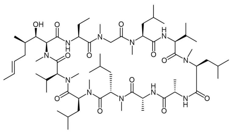
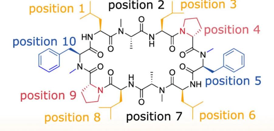
However, the oral delivery of Cyclosporine A has stimulated people’s enthusiasm for the development of oral peptides, but the key to completely opening the door to oral peptides has not been fully presented, and people are still searching for the realization of oral bioavailability of peptides. In addition to the stability of peptides and their resistance to degradation by a variety of enzymes, how to cross the cell membrane is also one of the focuses of research. Researchers from the University of ETH in Switzerland recently simulated the molecular dynamics of peptide crossing the cell membrane using a flexible decacyclic peptide (Fig.2).
In contrast to straight-chain peptides, which are too flexible, cyclic peptides have what are known as chameleon properties. Chameleons change skin color based on the environment, while cyclic peptides can change the molecular conformation based on the polarity of the environment. This property depends on the conversion of intramolecular and intermolecular hydrogen bonds in the cyclic peptide molecule.
In the so-called Open conformation of investigational peptide drugs, the hydrogen-bonded atoms of cyclic peptides are exposed to a polar environment (e.g., in blood or cytoplasm) to form hydrogen bonds with their counterparts in solvents, or with polar solvent molecules. But when the environment becomes non-polar (e.g., inside a cell membrane), cyclic peptides can instead adopt the so-called Closed conformation, wrapping themselves up and encapsulating themselves by forming intramolecular hydrogen bonds, encapsulating polar groups inside the molecule and exposing the non-polar structure to surface of the molecule, thereby reducing the polar area and the desolvation energy (the amount of energy required for the solute to be removed from the water or other solvents). This chameleon property of cyclic peptide is helpful in the development of oral peptide formulations because it can balance good water and fat solubility, which favors increasing cell permeability.
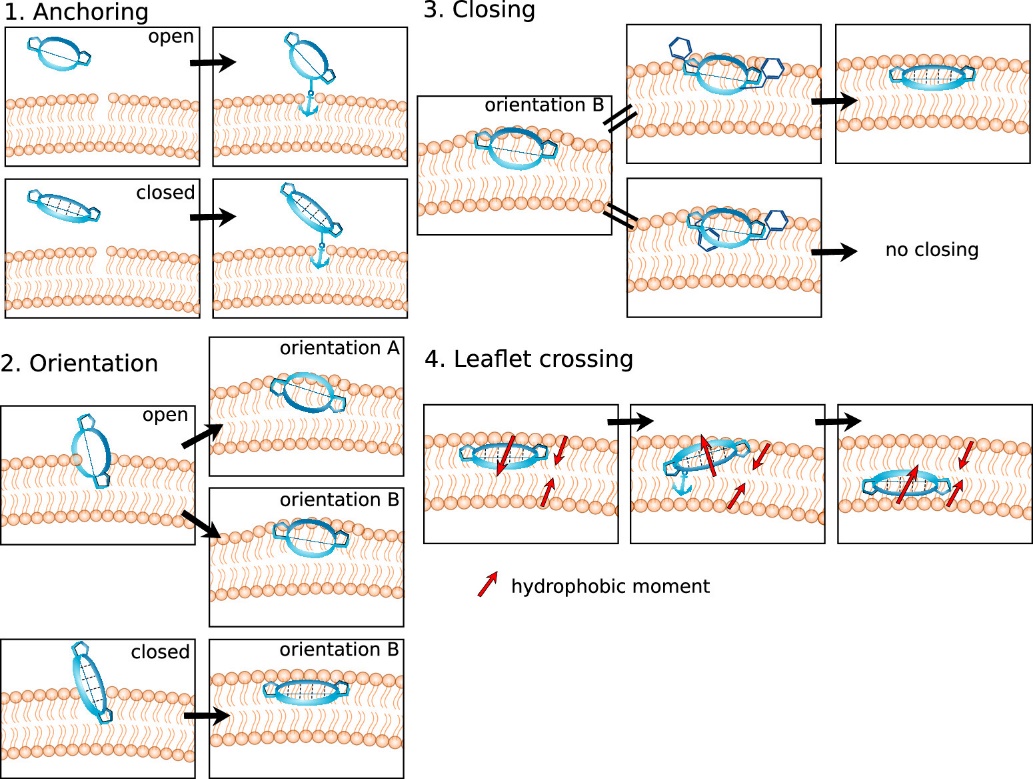
The Closed state is generally considered to be the predominant permeability state of cyclic peptides. However, it is a dynamic process that is controlled by a variety of factors such as sequence, molecular size, hydrophobic surface area, etc. Through molecular dynamics simulation, the researchers simulated the process of cyclic peptide molecules crossing the cell membrane through passive osmosis into the cell, highlighting the importance of flexible conformation for cyclic peptide membrane permeation, and dividing it into four steps (Fig.3):
- Specific peptide side-chain residues can act as molecular anchors to establish the initial contact of cyclic peptides with the cell membrane before the peptide molecule is inserted into the cell membrane.
- After anchoring to the cell membrane, the peptide molecule jumps directly into it, inserting itself into the interface between the polar head and non-polar tail regions of the cell membrane. In this process, the cyclic peptide exhibits two potential directions of arrangement, and the open state takes orientation A and B, and preference orientation A is preferred. Closed cyclic peptides take orientation B only.
- The Open state transitions to the Closed state, increasing the chance of it penetrating. At this time, the chameleon nature of the peptide molecules is very important for them to move from a polar environment to a non-polar environment. It is difficult for the cyclic peptide in the open state to achieve the subsequent diffusion process.
- Only cyclic peptide molecules in the closed state are likely to diffuse from this polar/non-polar mixed environment on the cell membrane surface through the non-polar lipid membrane leaflet and spread to the polar/non-polar interface on the other side. Through the anchoring and flipping mechanism, the final transmembrane journey is achieved.
Step 1
Simulated molecular dynamics showed that the anchor residues of the Open conformation and the Closed conformational cyclic peptide were different, due to the fact that the degree of contact of the residues was not the same in the two conformations. Given that the anchoring of amino acids is peptide conformation-dependent, characterizing the conformational behavior of cyclic peptides is critical for rational peptide drug design. Understanding the main conformations of peptides before they enter the cell membrane and the corresponding residue exposure will help optimize the design of membrane-permeable cyclic peptides. This mode of membrane permeability initiated by side chain anchoring of amino acid residues can be generalized to other molecular modalities other than peptides, such as increasing the membrane permeability of molecules by means of moderate aliphatic chain conjugation.
Step 2
The peptide is inserted into the membrane and parallels itself to the membrane plane. If the insertion occurs in a molecule with an open conformation, the peptide may take two directions of arrangement, A and B. However, if the peptide insertion is already in a closed conformation (pre-folded), only direction B is observed. This can also be useful for the design of other macrocyclic peptides, especially those with continuous hydrophobic surfaces. Only orientation B favors the conversion of peptides from the Open to Closed conformation.
Step 3
The change of conformation from the open state to the closed state is a critical process that has an important impact on the subsequent intramembrane diffusion. This conformational transition not only changes the spatial structure of the molecule, but also significantly affects its interaction with the membrane environment, thus determining the movement mode and diffusion efficiency of the molecule in the membrane.
Step 4
The peptide molecule that enters the cell membrane must expand to the other side of the membrane (lower leaflet). As the final step of crossing the membrane, the peptide molecule needs to go through a similar process as the first step, that is, its amino acid residues need to be anchored to the polar head of the lower leaflet, and this process also involves the reversal of the direction of the cyclic peptide, resulting in a change in the direction of the hydrophobic moment.
Simulations have shown that the effects of amino acids (e.g., leucine, phenylalanine, proline) may be highly correlated with the peptide sequence, depending on the position of the amino acid in the peptide as well as other residues. The goal of this molecular dynamics simulation of the cyclic peptide permeation pathway is to improve the bioavailability of peptides, especially oral bioavailability, and to develop more peptide drugs that can be delivered orally.
References
Linker, Stephanie M., et al., Lessons for Oral bioavailability: how Conformationally flexible cyclic peptides enter and cross lipid membranes. Journal of medicinal chemistry 66.4 (2023): 2773-2788.
]]>Protein-protein interactions (PPI) play a crucial role in various biological processes, and the misregulation of PPI is often associated with the onset of diseases. Peptides, especially linear peptides, face limitations such as susceptibility to proteolytic degradation and poor membrane permeability in practical applications. Therefore, modifications of peptides are often necessary. Peptide modifications can be categorized into N-terminal modification, C-terminal modification, side-chain modification, backbone modification, and cyclization. Among them, peptide cyclization is a common and effective way to enhance peptide stability and binding affinity.
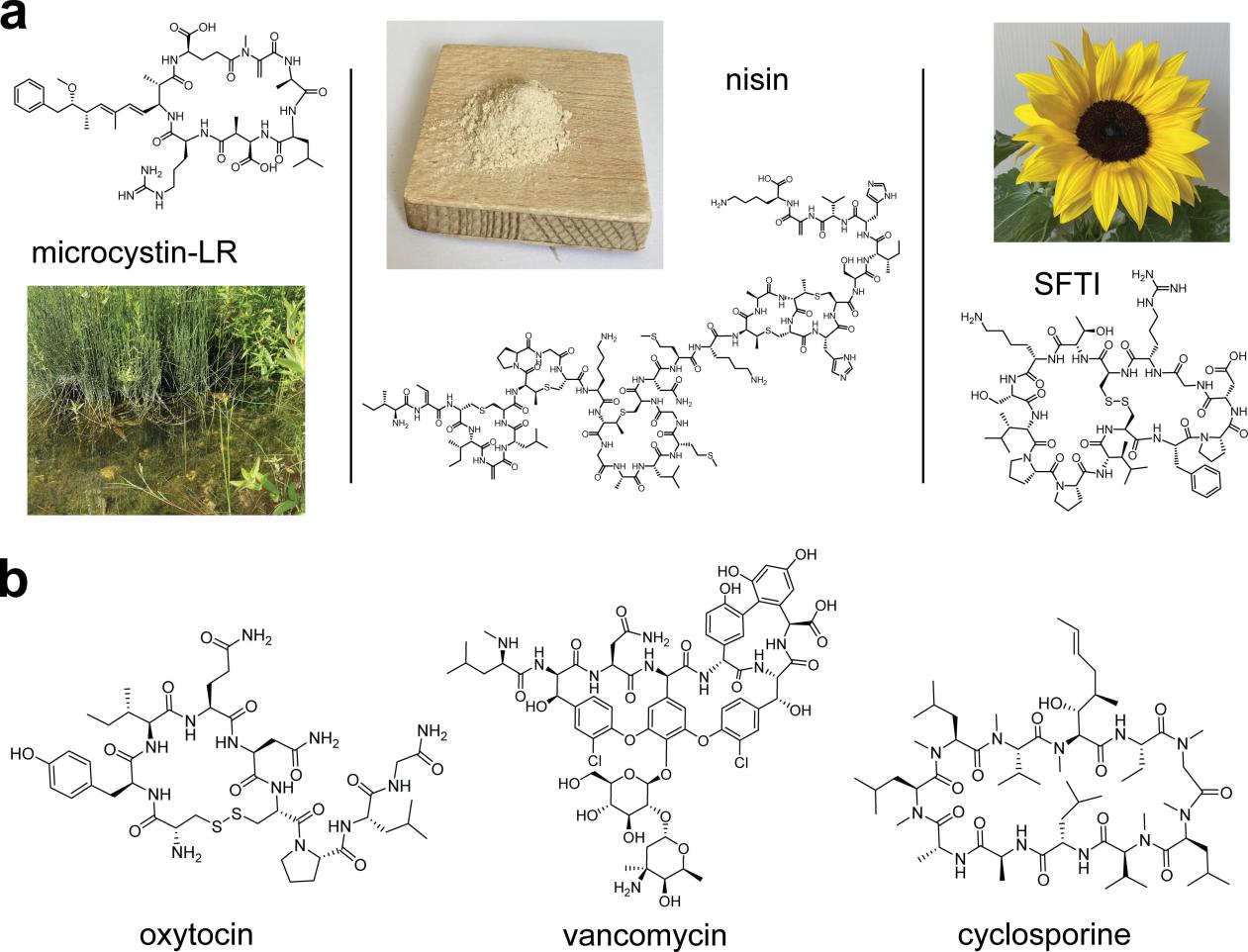
Many natural cyclic peptides or their derivatives are used in clinic therapeutics. For instance, romidepsin, a bicyclic peptide extracted from Chromobacterium violaceum, inhibits histone deacetylase and is clinically used as an anti-tumor drug for treating T-cell lymphoma. Sunflower trypsin inhibitor (SFTI) is the bicyclic peptide that found in seeds of sunflower. Voclosporin, a large cyclic peptide derived from cyclosporin, was approved in 2021 for the treatment of lupus nephritis (LN). Pasireotide, an analog of somatostatin, is used clinically to treat Cushing’s disease when surgery is ineffective.
Cyclic Peptides with Amide Bonds
Initially, researchers used natural amino acids like lysine (Lys), glutamic acid (Glu), or aspartic acid (Asp) residues to form intramolecular amide bonds for cyclic peptide synthesis. The Fmoc/t-butyl solid-phase synthesis method is commonly used today. In this method, the lysine side-chain amino group is protected by allyloxycarbonyl (Alloc), and the carboxyl group of Glu or Asp side chains is protected. After synthesizing the linear peptide chain through solid-phase synthesis, the protecting groups are removed using metal Pd, and the cyclization is performed using coupling agents such as PyBOP. A drawback of this method is the use of heavy metals like Pd during deprotection, which can be harsh.
Cyclic Peptides with Cysteine Residues
Utilizing cysteine residues to form intramolecular disulfide bonds is a common strategy for constructing large cyclic peptides. Various methods, such as air oxidation, iodine oxidation, potassium ferricyanide oxidation, dimethyl sulfoxide oxidation, and thallium trifluoroacetate oxidation, are employed to protect the thiol groups of cysteine residues when two or more disulfide bonds are present, using protective groups such as trityl (Trt), monomethoxytrityl (Mmt), acetylamidoethyl (Acm), t-butyl (t-Bu), benzyl (Bzl), or p-methoxybenzyl (Tmob) for thiol protection. When constructing cyclic peptides via disulfide bonds, an appropriate linker is required to enhance the affinity and membrane permeability of the cyclic peptide.
Stapled Peptides
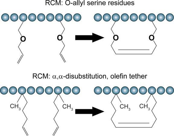
Stapled peptides involve cross-linking the side chains of any two amino acids in a peptide or cross-linking one amino acid side chain to the end of the peptide, forming a stable α-helix or β-fold conformation, primarily referring to α-helical peptides supported by olefinic or alkyne linkers. Since α-helices constitute 30% to 40% of ordered proteins and play a crucial role in the binding of many important biological receptor proteins, stapled peptides with α-helices exhibit higher target affinity, stronger cell penetration, and resistance to proteolytic degradation. The synthesis strategy for stapled peptides involves introducing non-natural amino acids with α-methyl or α-alkene into the solid-phase peptide chain and then cyclizing through ring-closing metathesis (RCM). In RCM reactions, non-natural amino acids are usually inserted at positions i, i+3, i+4, or i+7 in the peptide sequence.
Bicyclic Peptides
Naturally occurring cyclic peptides generally consist of no more than 10 amino acids, resulting in relatively small molecular masses. Such peptides are less susceptible to proteolytic degradation in the body, but they are poorly absorbed orally. On the other hand, cyclic peptides with more than 10 amino acids, although more structurally diverse and stable than linear peptides, still lack sufficient stability in vivo. To address the issue of poor stability of large cyclic peptides in vivo, researchers have turned to synthesizing bicyclic peptides. Compared to monocyclic peptides, bicyclic peptides have a more rigid structure, and with proper design, one ring is responsible for binding to cell surface proteins, providing them with targeting properties. The other ring, after the entire molecule is transported into the cell, binds to the target protein, exhibiting inhibitory activity.
Plecanatide is a peptide with a structure almost identical to human uroguanylin, consisting of 16 amino acids. Plecanatide, through two pairs of disulfide bonds, exhibits an active conformation at physiological pH 5.0 in the proximal duodenum and jejunum, binding to guanylate cyclase C (GC-C) receptors. It acts as a guanylate cyclase receptor agonist, promoting the excretion of urinary sodium, regulating acid-base ions in the gastrointestinal tract, inducing fluid transport into the gastrointestinal tract, and increasing gastrointestinal motility. Plecanatide is suitable for treating chronic idiopathic constipation in adults.
Conclusion and Prospects
The trend in peptide drug development has shifted from naturally isolated cyclic peptides to optimized cyclic peptide analogs to enhance peptide efficacy, stability, and pharmacokinetic properties. The application of cyclic peptides has also expanded to various areas, including disease treatment, drug targeting, imaging, and diagnostics. Current research on cyclic peptides still focuses on stability, and their drug-like properties require further investigation. Rational drug design could be based on specific disease targets, focusing on the binding sites of key amino acids. Subsequently, through biological activity evaluations, cyclic peptides with optimal drug-like properties may be discovered.
References:
- Ji, X., et al., Cyclic Peptides for Drug Development, Angew Chem. Int. Ed., 2023.
- Walensky, L. D., and Bird, G. H., Hydrocarbon-Stapled Peptides: Principles, Practice, and Progress, J Med Chem., 2014, 57(15): 6275-6288.
Peptides are a class of compounds formed by linking multiple amino acids through amide bonds, and their molecular size is between small molecule chemical drugs and macromolecular biological drugs. Compared with small molecule chemical drugs, peptides have higher efficacy, selectivity and specificity, and lower metabolic toxicity. Compared with macromolecular biological drugs, peptides have lower immunogenicity and can penetrate deeper into the target, and continuous production costs are lower. Based on these unique advantages, in recent years, peptide drugs have attracted more and more attention. According to statistics, the growth rate of the peptide drug market is twice that of other drugs. At present, a variety of peptide drugs have entered the market for the treatment of diabetes, osteoporosis, cancer, multiple sclerosis, chronic pain, HIV infection and many other diseases.
As we know, active peptides are the material basis for drug efficacy, so in the development process of peptide drugs, the quality control of active peptides (API) is very important, and the control of peptide-related impurities is an important work of quality control, impurities not only affect the quality and safety of clinical trial samples, in the early stage of drug discovery, These impurities may also affect the judgment of drug efficacy, and inadequate research on impurities may even lead to incorrect conclusions.
Classification of Impurities and Ways of Production
- Impurities related to the synthesis process
Chemical synthesis technology is the gold standard for peptide production. The process was initially carried out in solution, and later solid-phase peptide synthesis was introduced. After decades of continuous development, the technology has become very mature. The steps of chemically synthesizing the polypeptide include: hanging resin-deprotection group-activation and cross-linking-peptide cleavage. In this process, the steps of deprotection, activation and cross-linking are prone to impurities, which are usually points that require quality control.
Missing peptide: As the name suggests, a missing peptide is a peptide that is missing one or more of the required amino acids, and the transient protective group of amino acids that is not completely removed, insufficient activation of the amino acid to be bound, or low cross-linking efficiency can lead to the production of the missing peptide. The identification method of the missing peptide, that is, the mass difference between the impurity and the target peptide observed in the mass spectrometry is the molecular weight of the missing amino acid minus water.
Insertional peptide: During the cross-linking process of synthetic peptides, amino acids are usually fed in excess. If the excess amino acids are not fully washed or the reaction time is too long, the amino acids will be cross-linked again to generate insertion peptides. For the identification method of the inserted peptide, the mass difference between the impurity observed in the mass spectrum and the target peptide is the molecular weight of the inserted amino acid minus water.
Misfolded peptide: It can arise when both amino acid deletions and amino acid insertions occur during a solid-phase reaction process.
Covalently attached peptide impurities: After solid phase synthesis, incomplete removal of permanent protective groups will lead to covalent attachment of protective groups to the peptide sequence, resulting in covalent attached peptide impurities. For the identification method of such impurities, if the molecular weight of the impurity in the mass spectrum differs from the molecular weight of the target peptide by 56 Da, then the attached protective group is the tBu protective group, if the molecular weight of the impurity in the mass spectrum differs from the molecular weight of the target peptide by 100 Da, then the attached protective group is the tBoc-TCS protective group.
- Degradation related impurities
Degradation related impurities include impurities formed by degradation mechanisms during manufacturing and impurities formed by degradation mechanisms during storage. The factors that cause the instability of peptide include oxidation, light, high temperature, pH, ionic strength change and adsorption, etc. The amino acid type and the position of amino acid in the peptide chain have important effects on the stability of peptide.
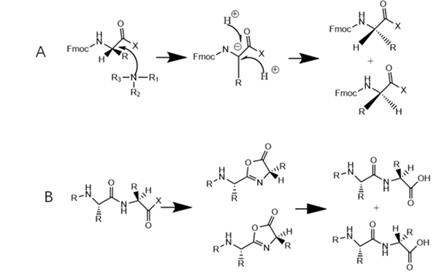
Epipeptide: it refers to the impurity formed by amino acid residues with one or more unexpected chiral configurations in the sequence, which may be derived from optical isomers in the starting materials, or amino acids during the synthesis by enolization or azalactonization of the chiral center. The chromatographic behavior of epi-peptide impurities is very close to that of the main component, and the molecular weight is also the same as that of the main component, so separation and identification are more difficult. Deuterated reagents can be used to hydrolyze and derivatize peptides, and the amino acids that are prone to racemization in the peptide chain can be judged by the content of chiral isomers of amino acids obtained from hydrolysis, and possible epimer peptides can be prepared by directional preparation. targeted research.
Broken peptide: Peptide bonds may be broken when peptide encounter high temperature or pH changes, resulting in broken peptides. Studies have shown that alkaline conditions are most likely to cause peptide bond breaks. The identification of the broken peptide is relatively simple, and which amide bond is broken can be inferred from the molecular weight difference between the impurity and the parent peptide.
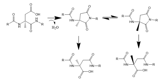
Deamidation impurities: When the peptide contains glutamine or aspartate residues, the amide bond may be removed to form glutamic acid or aspartate residues, which can be formed by two mechanisms, one is direct hydrolysis (easy to occur at pH<5), and the other is through β transformation (easy to occur under alkaline conditions), which attacks the carbonyl group on the side chain through the N atom of the amide bond in the main chain. Form a succinimide ring (a type of azalactone), which hydrolyzes under alkaline or neutral conditions to form a carboxyl group. The succinimide hydrolysis process is often accompanied by two side reactions, namely isomerization and racemization, and finally form four isomers with the same molecular weight. The identification of deamidation impurities is mainly through mass spectrometry, and the impurities detected with molecular weight +1 Da are usually deamidation impurities.
Isomerized impurity: Aspartic acid is an amino acid that is prone to isomerization. Aspartic acid is easily cyclized to the main chain due to dehydration or ammonia in the side chain to form succinimide. Succinimide has poor stability and is easy to hydrolyze. In the process of ring opening, isomers can be formed, and epi-peptide by-products can also be produced at the same time, and its formation mechanism is similar to that of asparagine deamidation.
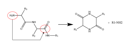
Diazinoperone and pyroglutamate: Diazinoperone refers to the N-terminal active group (usually an amino group) attacking the carbonyl group of the amide bond in the β-site main chain to form diketopiperazine analogens, resulting in a missing peptide with two amino acids, and the reaction is more likely to occur when the N-terminal amino acid hindrance is small. Pyroglutamic acid refers to the N-terminal containing glutamic acid which is cyclized to form pyrrolidone carboxylic acid. The impurity can be identified by mass spectrometry with a molecular weight of 17Da less than the parent peptide (NH3 removed).
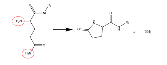
Polymer: Intermolecular aggregation and self-association of peptides can lead to the formation of high molecular weight impurities known as polymers. There are two types of polymerization mechanisms: covalent and non-covalent. Non-covalent polymerization is formed by electrostatic or hydrophobic interaction, which is an intermediate state. When the solution concentration, pH or ionic strength changes, this force is easily weakened or disappears, and the polymerization state returns to the monomer state. when the peptide chain contains thiol or tyrosine, it can cause covalent polymerization through the formation of disulfide bonds or the formation of dityrosine. Covalent polymerization is a stable polymerization state that does not change with peptide concentration or pH, ionic strength, etc. Due to the further increase of molecular weight and volume of polymer impurities, it is easy to increase the immunogenicity and cause allergic reaction, so the content of polymer should be controlled reasonably. Polymers are usually detected by volumetric exclusion chromatography, which can be used in conjunction with mass spectrometry to determine the multiple of their polymers.
Oxidative impurities: Oxidative impurities are formed when peptides are exposed to oxygen and light during storage. Amino acids that are prone to oxidation include sulfo-containing amino acids, histidine, tyrosine, and tryptophan.
Peptide-related impurities are not only a problem that needs continuous attention in the GMP supervision during the drug development and production process, but also may cause misjudgment of developers in the early stage of drug discovery and development because of the antagonism of impurities or the super potency of the drug. Therefore, it is usually recommended to use peptides from different sources for retesting when carrying out biological tests to eliminate the influence of impurities on the drug efficacy.
References
- Muttenthaler M, King GF, Adams DJ, Alewood PF, Trends in peptide drug discovery. Nat Rev Drug Discov 20:309-325.
- V.Sanz-Nebot, I.Toro, Investigation of synthetic peptide hormones by liquid chromatography coupled to pneumatically assisted electrospray ionization msaa spectrometry:analysis of a synthesis crude of peptide triptorelin, Rapid Commun. Mass Spectrom,2001;15:1031-1039.
- Matthias D’Hondt, Nathalie Bracke, Related impurities in peptide medicines, Journal of Pharmaceutical and Biomedical Analysis 101 (2014) 2–30.
- Shikha Patel, Vivek K. Vyas, Priti J. Mehta, A Review on Forced Degradation Strategies to Establish the Stability of Therapeutic Peptide Formulation, International Journal of Peptide Research and Therapeutics (2023) 29:22.
- Brian Gregg,Aleksander Swietlow,Control Strategies for Synthetic Therapeutic Peptide APIs Part III: Manufacturing Process Considerations.
- Virender K. Sharma a& Nigel J.D. Graham , Oxidation of Amino Acids, Peptides and Proteins by Ozone: A Review, Ozone: Science & Engineering, 32: 81–90.
In recent years, more and more studies have described the antioxidant capacity of plant protein-derived peptides. Plant proteins are considered a new source of antioxidant peptides that can not only save energy, but also enhance the treatment of oxidation-related diseases, delay the oxidation of food, and thus improve the quality of life. Despite an increasing number of reports on the preparation, structure, and activity of antioxidant peptides, and the commercialization of some antioxidant peptides, a systematic review and evaluation of plant protein-derived antioxidant peptides is still lacking. This article summarizes the latest advances in plant protein-derived antioxidant peptides, including (1) the preparation, purification, identification, and application of plant protein-derived antioxidant peptides by enzymatic hydrolysis; (2) Influencing factors, evaluation methods, and mechanism of antioxidant protein hydrolysates or peptides.
Traditional purification and identification methods of antioxidant peptides are expensive and time-consuming. To solve this problem, researchers have conducted extensive exploration and research on computer-aided mathematical models (molecular docking, QSAR studies, etc.), bioinformatics tools, and real-time updated protein databases. In addition, the authors of the reviews believe that future research should focus on the large-scale commercial production of plant protein-derived antioxidant peptides, comprehensive studies on the structure-activity relationship of antioxidant peptides, an in-depth understanding of antioxidant mechanisms, and confirmation of their health benefits in vivo.
What is the plant protein-derived antioxidant peptide?
Antioxidant peptides are specific protein fragments possessing antioxidant activity and antioxidant peptides derived from plant protein are called plant protein-derived antioxidant peptides. With increasing consumer demand for high nutritional and functional proteins, researchers are shifting their focus from traditional raw proteins to plant-based proteins. In particular, natural antioxidant peptides derived from plant proteins have attracted worldwide attention due to their advantages of environmental protection, sustainability, low cost, and non-toxic side effects.
Evaluation methods and mechanism of antioxidant peptides
The effective prevention of oxidative stress and food oxidation of antioxidant peptides attracted researchers to evaluate their antioxidant capacity. At present, there are three methods to evaluate the antioxidant capacity of peptides: chemical method, cell biology method in vitro, and animal experiment in vivo. The potential mechanisms of antioxidant peptides are as follows: (1) direct scavenging of reactive oxygen species; (2) chelated metal ions; (3) Inhibition of lipid peroxidation; (4) Activate the body’s antioxidant defense system.
Enzymatic preparation of plant protein-derived antioxidant peptides
From the point of view of sustainable development and utilization of biological resources, protein resources in the by-products or wastes produced during plant processing have great potential to produce bioactive peptides. The rational development and utilization of plant processing by-products or wastes are beneficial to improve the nutritional utilization value of wastes, save energy and protect the environment. In particular, wheat gluten, wheat germ, rapeseed, watermelon seed, corn gluten, rice bran, oat bran, palm kernel cake, and wine-making grains have been used to produce antioxidant peptides.
Plant proteases (animal proteases, plant proteases, and microbial proteases) are hydrolyzed first to form proteolysis, followed by further purification, membrane separation, electrophoresis, chromatography (gel permeation chromatography (GPC), ion exchange chromatography (IEC), reversed-phase high-performance liquid chromatography (RP-HPLC), etc.) to obtain antioxidant peptides. The obtained antioxidant peptides were identified by mass spectrometry or mass spectrometry/mass spectrometry, and their antioxidant activities were evaluated by in vitro and in vivo experiments. At present, the main preparation methods for peptides are chemical extraction, fermentation, synthesis, and enzymolysis.
Purification and identification of plant protein-derived antioxidant peptide
The enzymolysis product of proteins is a mixture consisting of partially unhydrolyzed proteins, peptides with different chain lengths, hydrophobicity and net charge, and free amino acids. To evaluate the structure and activity of antioxidant peptides more accurately, it is necessary to develop suitable purification methods. At present, the main separation and purification technologies of peptides include chromatography (GPC, IEC, RP-HPLC), membrane separation, and a combination of these technologies. Identification of protein hydrolysates is usually accomplished by mass spectrometry, mass spectrometry/mass spectrometry, and bioinformatics techniques.
After a series of isolation and purification schemes, the peptide structure needs to be identified. DNA translation, mass spectrometry, and peptide sequencing are commonly used to identify peptides. It is worth noting that mass spectrometry has been widely used in the identification of peptide sequences because of its high efficiency, sensitivity, and good reproducibility. In addition, several online databases have become popular for researching antioxidant peptides. Table 2 summarizes the main bioinformatics analysis tools.
Factors influencing the action of antioxidant peptides
It is important to summarize the properties and activities of plant-derived antioxidant peptides as comprehensively as possible for guiding and predicting the relationship between the structure and function of plant-derived antioxidant peptides. The activity of antioxidant peptides is mainly affected by amino acid composition, sequence, and molecular weight.
Analysis of the structure-activity relationship of antioxidant peptides by computer-aided molecular modeling
QSAR is a mathematical model to establish quantitative dependence between the structure-activity relationships of a series of compounds. QSAR is used to evaluate the correlation between the molecular structure and activity of antioxidant peptides, which provides a good theoretical basis for the prediction, isolation, and purification of peptide activity and elucidation of molecular mechanism.
Future research direction and existing problems
As mentioned earlier, proteins derived from plant products can be used to produce antioxidant peptides. On this basis, the research progress of plant-derived antioxidant peptides was reviewed in this paper. The molecular mechanism of these peptides at the level of the whole gene will be verified with the advancement of science and technology in the future and the continuous investment in the research of these peptides in vivo. In addition, the preparation of nanoparticles and microcapsules of antioxidant peptides and their synergies with other active ingredients are also the focus of future research. Future research and industrial cooperation will help achieve the long-term goal of commercializing plant antioxidant peptides.
]]>Antiviral drugs are a class of drugs used to treat viral infections. While most antiviral drugs target specific viruses, broad-spectrum antivirals are effective against multiple viruses. Most of the antiviral drugs available nowadays target HIV, herpes, hepatitis B and C viruses, and influenza A and B viruses. The coronavirus represented by COVID-19 has also become the focus of antiviral drug development.
Peptide and peptidomimetic antiviral drugs
The generic names of many antiviral drugs end in “vir” and the chemical structure of these antiviral drugs are in the form of peptidomimetics, such as atazanavir, lopinavir, nelfinavir, boceprevir, telaprevir, nematvir/ritonavir, not to mention antiviral drugs ending with “tide” including bulevirtide.
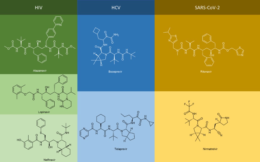
The use of peptidomimetics as a therapeutic agent has many advantages compared with peptide drugs, e.g., enhanced binding force, improved metabolic stability, and good bioavailability. It is an important means for the development of antiviral drugs of this kind to transform traditional peptide drugs into peptidomimetics through different strategies of chemical modification, to further improve receptor affinity and pharmacokinetic properties.
Common strategies for peptide modifications
Traditional peptide drugs are hampered by poor metabolic stability and low oral bioavailability, therefore, researchers use chemical modifications to enhance their stability and bioavailability. Chemical modifications of peptides generally include the following methods:
1. N-Terminal modifications/C-Terminal modifications
The minimum action of chemical modification is to block the N-terminal of a peptide with an acetyl group and modify the C-terminal into an amide. Such modifications can reduce the Nα-mediated degradation of DKP (diketopiperazine). This protective effect is particularly significant for peptides with smaller molecular weights. By the end of 2021, among the 62 marketed short peptides (consisting of 15 amino acid residues or less), 37 peptides are modified at their N-terminal by acylation, pyroglutamate, or cyclization, and only 9 linear peptides have free Nα groups. Terminal modifications have a significant effect on the secondary structure of the peptide because it deprives the molecule of its electrical charge. The change in the spatial structure of the peptide, in turn, can affect molecular stability.
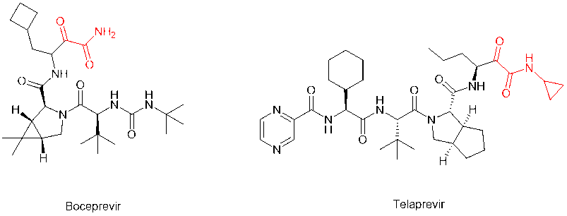
In addition to the two most common modifications above, the alpha-keto amide (alpha-keto-carbonyl amide) structure can be loaded on the C-terminal to introduce the keto-carbonyl group into the peptide molecule and transform it into a covalent peptide-like drug that can form a covalent bond with the receptor. For example, peptidomimetic drugs boceprevir and telaprevir, which target the serine protease of HCV virus, have alpha-keto amide in their C-terminus. The modification greatly improves the potency and pharmacokinetic properties of the parent compound.
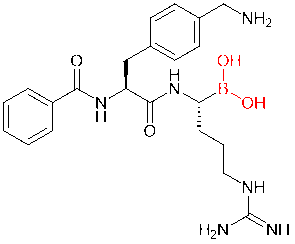
Boric acid structures, another increasingly popular covalent drug, are also frequently introduced into peptide molecules. In a study, for example, scientists added boric acid structures to dipeptide inhibitors of Zika, West Nile, and dengue virus proteases, replacing the carboxylic acid or amide groups. As a result, the derived boric acid inhibitor had a thousandfold increase in affinity.
2. Bioisostereric replacement of amide
The skeleton of the peptide is composed of amide bonds, which is very important for the physical and chemical properties of the peptide. Replacing amide bonds by bioisostere is a vital strategy for researchers to move from peptide forward to peptidomimetics. Common amide electron isoplatelets include esters, thioamides, 1,2,3-triazoles, 1,2,3-triazoles, sulfonamides, phosphoramidites, hydroxy ethylene, hydroxy ethylamine, etc.
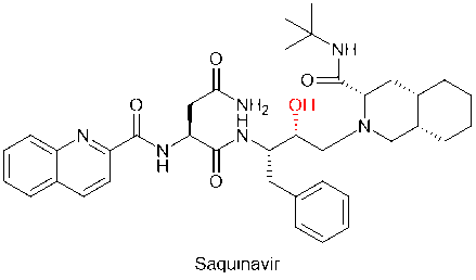
Practices of chemical modification methods that replace amide bonds by bioisostereric replacement are numerous in peptidomimetic drug development. For example, saquinavir, the first protease inhibitor against HIV, replaced the unstable amide bond in the peptide main chain with the hydroxymethyl structure, improving the metabolic stability of the drug.
Amino acid modifications are extremely extensive, including the substitution of non-proteinogenic amino acids for protein amino acids, such as D-amino acids for L-amino acids, thus improving the hydrolytic stability of the substrate. Researchers have a growing library of non-protein amino acids that can support different types of amino acid modification.
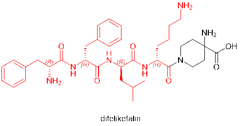
D-amino acids have even become regular residues of peptide drugs in the market. Among the peptide drugs approved in 2021 are voclosporin (D-Ala) and odevixibat (D-4-hydroxyphenylalanine), which contain D-amino acids. Furthermore, defelikefalin (korsuva, a pain-relieving opioid peptide for moderate to severe itching), approved by the FDA in 2021, uses a full D-amino acid (D-Phe, D-Leu, D-Lys) strategy.
4. Cyclic Peptides
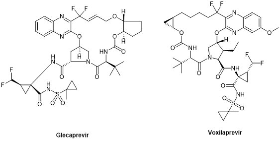
Peptide cyclization has always been an important chemical modification strategy for peptides. Since cyclosporin A, the first oral peptide preparation, was released in the 1980s, peptide cyclization has been regarded as one of the key ways to improve the stability, pharmacokinetic properties, and bioavailability of peptides. By 2021, 47% of the nearly 105 peptide therapeutic agents and diagnostic reagents approved by the FDA, EMA, and FMDA are cyclic peptides, of which 37% are single-cyclic peptides and 10% are polycyclic peptides. Cyclic peptides have also been found in anti-HCV drugs, especially in large cyclic reversible NS3/4A serine protease inhibitors, such as simeprevir, glecaprevir, and voxilaprevir.
R&D of Antiviral Peptides
1. Anti-SARS-CoV-2 virus peptidomimetic drugs
Perhaps the most high-profile example of the peptidomimetic antiviral therapy is Paxlovid, which helped stabilize the global COVID-19 outbreak caused by the SARS-CoV-2 virus. Paxlovid is an FDC (Fixed-dose combination) that contains two active substances, nirmatrelvir and ritonavir, which, as mentioned above, are peptidomimetics. Ritonavir was marketed as an anti-HIV drug but was quickly replaced by a new generation due to its potency and side effects. However, due to its high binding capacity with CYP3A4 enzyme and its inhibition of CYP3A4 mono plus oxidase (through different mechanisms, including (I) the formation of the metabolic intermediate complex (MIC) that closely bound to the heme group; (II) strong binding of unmodified ritonavir to heme iron; (III) heme destruction; (IV) covalent linking of active ritonavir intermediates to CYP3A4 deco-basing protein, etc.), ritonavir acts as a pharmacokinetic enhancer in Paxlovid.
Through its inhibition of CYP3A4, nirmatrelvir, the true antiviral active substance, is protected from enzymatic degradation of CYP3A4, increasing its half-life and drug exposure. The pharmacological action of nirmatrelvir is its inhibition of 3C-like protease (3CLPRO, also known as Mpro). Nirmatrelvir is therefore a Mpro protease inhibitor. Mpro is a cysteine protease whose cleavage of the S-glycoprotein on the spike protein of SARS-CoV-2 is critical to the biological process by which the virus fuses with host cells and infects them. Therefore, its inhibition is central to the pharmacological efficacy of Paxlovid.
Nirmatrelvir is a covalent peptidomimetic drug. Based on the non-covalent binding pocket to the receptor protein, the core building block of nirmatrelvir (the cyano group warhead), is the active electrophilic group on the covalent drug. It can interact with the sulfhydryl group of Cys145 residues on the Mpro protease to form thioimimate, which can bind the targeted receptor protein closely through covalently, and achieve high efficacy under the condition of a lower dose. It is important to note that Paxlovid is an oral antiviral based on the pharmacokinetic profile of the pharmacokinetic enhancer ritonavir and nirmatrelvir itself.

2. Anti-HCV peptidomimetic drugs
Anti-HCV (hepatitis C virus) peptide drugs are also one of the core tasks. Hepatitis C is a liver disease caused by the hepatitis C virus (HCV). Currently, treatment for HCV infection is based on direct antiviral drugs (DAAs) that target specific non-structural proteins and disrupt viral replication.
In 2011, the FDA approved first-generation DAAs, boceprevir and telaprevir, which target NS3/4A in patients with genotype 1 HCV infection and both contain alpha-keto amide in their C-terminus. These DAAs are not cheap to use in combination with pegylated interferon (peg-IFN) and ribavirin.
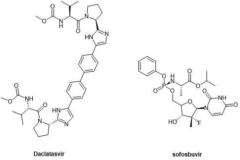
The second generation of DAAs, peptide-like drugs daclatasvir and sofosbuvir, which were introduced as NS5A and NS5B inhibitors, respectively, represent a breakthrough in HCV treatment, improving sustained viral response rate (SVR) in more patients.
Glecaprevir and voxilaprevir, the third generation of DAAs developed for NS3/4A, target all genotypes, including genotype 3 that represents the milestone in HCV treatment, and are cyclic peptides.
3. Anti-HIV peptidomimetic drugs
Anti-HIV drugs can be divided into nucleoside reverse transcriptase inhibitors (NRTIs), non-nucleoside reverse transcriptase inhibitors (NNRTIs), protease inhibitors (PIs), fusion inhibitors, CCR5 antagonists, integrase strand transfer inhibitors (INSTIs), attachment inhibitors, post-attachment inhibitors, and many other types. Among protease inhibitors, many drugs are peptidomimetics.
Saquinavir, a peptidomimetic drug approved by the FDA in 1995, was one of the first-generation protease inhibitors which were based on extranets such as hydroxy ethylene and hydroxy ethylamine electrons. The central hydroxyl group was bound to catalytic aspartic acid residues by simulating the transition state of the hydrolysis step. Ritonavir was also one of the first-generation anti-HIV protease inhibitors.
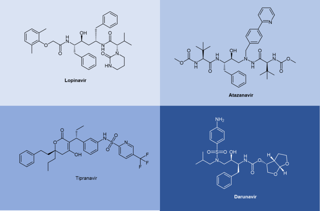
Lopinavir is a second-generation anti-HIV protease inhibitor developed by Abbott to improve the properties of ritonavir. Lopinavir cannot be administered alone and thus became the first compound preparation to be used in combination with ritonavir as a pharmacokinetic enhancer. Atazanavir was approved in 2003, and it became the first protease inhibitor to be effectively administered once a day. Its half-life is longer than that of previous protease inhibitors. Tipranavir was approved in 2005 and expanded for pediatric use in 2008. Because of many differences in its structure, it maintains effectiveness against certain strains of drug-resistant HIV and appears to have a higher genetic barrier, requiring many mutations to develop resistance. Darunavir, approved by the FDA in 2006, represents the latest anti-HIV protease inhibitor.
Summary
Peptidomimetics are powerful tools in pharmaceutical chemistry. They transform active peptides into complexes that combine the characteristics of peptides and small molecule drugs, significantly improving their pharmacological and pharmacokinetic properties over peptide drugs, and can be delivered orally on a large scale. The past decade witnessed a growing interest in antiviral peptides, and the emergence of Paxlovid demonstrated the tremendous value of antiviral peptides. With the deepening understanding of the pathogenic process of viruses, the combination of different antiviral drugs may become a reality, and the development of broad-spectrum antiviral drugs is also the research direction of scientists. It’s believed that the new generation of antiviral drugs ending with “vir” will continue to improve in efficacy, safety, resistance to drug resistance, delivery methods, and many other areas.
References
- Rossignol JF Nitazoxanide: a first-in-class broad-spectrum antiviral agent. Antiviral Res. 2014, 110: 94–103.
- Rick Daniels; Leslie H. Nicoll. “Pharmacology – Nursing Management”. Contemporary Medical-Surgical Nursing. Cengage Learning, 2011. p. 397.
- Kausar S, et al. A review: Mechanism of action of antiviral drugs. International Journal of Immunopathology and Pharmacology. 202135: 20587384211002621.
- Yang, Y. Side Reactions in Peptide Synthesis. Elsevier. 2015, 22-28.
- King, T.A. et al. (2021) Photocatalytic methods for amino acid modification. Chem. Soc. Rev. 50, 39–57.
- Robello, M. et al. The Alpha Keto Amide Moiety as a Privileged Motif in Medicinal Chemistry: Current Insights and Emerging Opportunities. J. Med. Chem. 2021, 64, 7, 3508–3545.
- Nitshe, C. et al. Peptide–Boronic Acid Inhibitors of Flaviviral Proteases: Medicinal Chemistry and Structural Biology. J. Med. Chem. 2017, 60, 1, 511–516.
- Kumari, S. et al. Amide Bond Bioisosteres: Strategies, Synthesis, and Successes. J. Med. Chem. 2020, 63, 21, 12290–12358.
- Ghosh, A.K. et al. Recent progress in the development of HIV-1 protease inhibitors for the treatment of HIV/AIDS. 2016, J. Med. Chem. 59, 5172–5208.
- Allerton, C. Pain Therapeutics: Current and Future Treatment Paradigms. Royal Society of Chemistry. 2013, pp. 56
- Nageswara, D. et al. (2021) Discovery of quinoxaline-based P1–P3 macrocyclic NS3/4A protease inhibitors with potent activity against drug-resistant hepatitis C virus variants. J. Med. Chem. 64, 11972–11989.
- Loos, N, H. et al. The Mechanism-Based Inactivation of CYP3A4 by Ritonavir: What Mechanism? Int. J. Mol. Sci. 2022, 23, 9866.
- Kjellin, M. et al. The effect of the first-generation HCV-protease inhibitors boceprevir and telaprevir and the relation to baseline NS3 resistance mutations in genotype 1: experience from a small Swedish cohort. Ups. J. Med. Sci. 2018, 123 (1), 50−56.
- Wei, L. et al. Long-term outcomes in patients with chronic hepatitis C in the current era of direct-acting antiviral agents. Expert Rev. Anti Infect Ther. 2019, 17 (5), 311−325.
- Ghosh, A. K. et al. The FDA Approved HIV-1 Protease Inhibitors for Treatment of HIV/AIDS. In Burger’s Medicinal Chemistry and Drug Discovery, 7th ed.; Abraham, D. J., Rotella, D. P., Eds.; John Wiley & Sons: Hoboken, NJ, 2010; Vol. 7, pp 1−74.
- Bold, G.; et al. New aza-dipeptide analogs as potent and orally absorbed HIV-1 protease inhibitors: candidates for clinical development. J. Med. Chem. 1998, 41, 3387−3401.
- Doyon, L.; et al. Selection and characterization of HIV-1 showing reduced susceptibility to the non-peptidic protease inhibitor tipranavir. Antiviral Res. 2005, 68, 27−35.
Summary of peptides for gastrointestinal studies
1. Motilin
Motilin is a 22-amino-acid hormone, released by the enteroendocrine M cells of the duodenum and proximal jejunum.
| Name | CAS | Sequence |
| Motilin, canine | 85490-53-5 | FVPIFTHSELQKIREKERNKGQ |
| Motilin (26-47), human, porcine | 52906-92-0 | FVPIFTYGELQRMQEKERNKGQ |
2. Gastrin
Gastrin is produced by G cells located in the gastric antrum and is the primary endocrine regulator of the secretory phase of a protein meal. The main function of gastrin is to stimulate gastric acid secretion, promote gastrointestinal motility, and participate in the maintenance of iron homeostasis. At the same time, gastrin can also stimulate the secretion of the pancreas, bile, and intestinal juice, and further decompose the food in the small intestine, which is beneficial to the absorption of nutrients in the small intestine.
| Name | CAS | Sequence |
| Gastrin I (human) | 10047-33-3 | Pyr–GPWLEEEEEAYGWMDF-NH2 |
| [Leu15]-Gastrin I (human) | 39024-57-2 | Pyr-GPWLEEEEEAYGWLDF |
| Gastrin I (1-14), human | 100940-57-6 | Glp-GPWLEEEEEAYGW |
| CCK-4 Acetate | 35144-91-3 | Trp-Met-Asp-Phe-NH2 |
| Pentagastrin | 5534-95-2 | Boc-β-Ala-Trp-Met-Asp-Phe-NH2 |
| Mini Gastrin I, human | 54405-27-5 | LEEEEEAYGWMDF-NH2 |
| Big Gastrin-1, human | 60675-77-6 | Pyr-LGPQGPPXLVADPSKKQGPWLEEEEEEAYGWMDF-NH2 |
| Gastrin I rat | 81123-06-0 | XRPPMEEEEEAYGWMDF |
3. Bombesin
Bombesin (BBS) is a 14-amino-acid peptide first isolated from the skin of the frog, Bombina bombina, in 1970 by Erspamer. It stimulates the release of gastrin and pancreatic enzymes and causes contraction of the gallbladder.
| Name | CAS | Sequence |
| Bombesin | 31362-50-2 | Glp-QRLGNQWAVGHLM-NH2 |
| Bombesin nonapeptide | 55750-00-0 | NQWAVGHLM |
4. Tachykinin
The tachykinins are a family of small peptides sharing a common C-terminal sequence, Phe-Xaa-Gly-Leu-Met-NH2, whose main members are SP, neurokinin A, and neurokinin B.
| Name | CAS | Sequence |
| Eledoisin | 69-25-0 | XPSKDAFIGLM |
| Kassinin | 63968-82-1 | DVPKSDQFVGLM |
| Neurokinin A | 86933-74-6 | HKTDSFVGLM |
| Neurokinin A(4-10) | 97559-35-8 | DSFVGLM-NH2 |
| Neurokinin B | 86933-75-7 | DMHDFFVGLM-NH2 |
| Substance P (1-7) | 68060-49-1 | RPKPQQF |
5. Somatostatin
Somatostatin belongs to the expanding family of small regulatory peptides which exert a variety of actions in different organ systems throughout the body.
| Name | CAS | Sequence |
| Octreotide | 83150-76-9 | FCFWKTCT (Disulfide bridge: Cys2-Cys7) |
| Octreotide Acetate | 79517-01-4 | FCFWKTCT |
6. Vasoactive intestinal peptide (VIP) and related peptides
The vasoactive intestinal polypeptide (VIP) is a member of a family of homologous neuro- and endocrine peptides which has three separate groups distinguished by the receptors which recognize them.
| Name | CAS | Sequence |
| Prepro VIP (111-122), human | 123025-94-5 | VSSNISEDPVPV |
| Prepro VIP (81-122), human | 111366-38-2 | HADGVFTSDFSKLLGQLSAKKYLESLMGKRVSSNISEDPVPV |
| VIP(6-28)(human, rat, porcine, bovine) | 69698-54-0 | FTDNYTRLRKQMAVKKYLNSILN-NH2 |
| [D-p-Cl-Phe6,Leu17]-VIP | 102805-45-8 | HSDAVXXDNYXRLRKQLAVKKYLNSXLN |
7. Neurotensin
Neurotensin is a 13–amino acid peptide originally detected in the bovine hypothalamus. Neurotensin can protect the airway from gastric mucosal injury caused by cold stimulation and can inhibit gastric acid secretion.
| Name | CAS | Sequence |
| Neurotensin(8-13) | 60482-95-3 | RRPYIL |
| Neurotensin | 39379-15-2 | Pyr-LYENKPRRPYIL |
| [Gln4]-Neurotensin | 61445-54-3 | Pyr-LYQNKPRRPYIL |
| Kinetensin | 103131-69-7 | IARRHPYFL |
| Xenopsin | 51827-01-1 | Glp-GKRPWIL |
8. Cholecystokinin
Cholecystokinin is secreted by cells of the upper small intestine. Its secretion is stimulated by the introduction of hydrochloric acid, amino acids, or fatty acids into the stomach or duodenum.
| Name | CAS | Sequence |
| Cholecystokinin Octapeptide (desulfated) | 25679-24-7 | DYMGWMDP |
| CCK-4 Acetate | 35144-91-3 | Trp-Met-Asp-Phe-NH2 |
9. Secretin
Secretin is a gastrointestinal peptide and a member of the pituitary adenylate cyclase-activating polypeptide (PACAP)/glucagon superfamily Sherwood et al.
| Name | CAS | Sequence |
| Secretin (28-54), human | 108153-74-8 | HSDGTFTSELSRLREGARLQRLLQGLV-NH2 |
| Secretin Acetate | 10813-74-8 | HSDGTFTSELSRLRDSARLQRLLQGLV-NH2 |
| Secretin (33-59), rat | 121028-49-7 | HSDGTFTSELSRLQDSARLQRLLQGLV-NH2 |
| Secretin, canine | 110786-77-1 | HSDGTFTSELSRLRESARLQRLLQGLV-NH2 |
| Secretin (5-27), porcine | 19665-15-7 | TFTSELSRLRDSARLQRLLQGLV-NH2 |
10. Pancreatic polypeptide
Pancreatic polypeptide (PP) is a 36–amino acid, 4.2-kDa polypeptide secreted by the islet F cells. PP belongs to the peptide YY/neuropeptide Y family of polypeptides.
| Name | CAS | Sequence |
| Pancreatic Polypeptide, bovine | 179986-89-1 | APLEPEYPGDNATPEQMAQYAAELRRYINMLTRPRY-NH2 |
| Pancreatic Polypeptide, human | 75976-10-2 | APLEPVYPGDNATPEQMAQYAADLRRYINMLTRPRY-NH2 |
| Pancreatic Polypeptide, rat | 90419-12-8 | APLEPMYPGDYATHEQRAQYETQLRRYINTLTRPRY-NH2 |
11. Peptide YY
Peptide YY (PYY) is a 36-amino-acid gut hormone released by L cells of the ileum/colon in proportion to ingested calories. PYY is functionally related to NPY and binds to the Y family of receptors, Y1R–Y6R.
| Name | CAS | Sequence |
| Peptide YY (3-36) | 126339-09-1 | IKPEAPGEDASPEELNRYYASLRHYLNLVTRQRY-NH2 |
| Peptide YY (3-36) Human | 123583-37-9 | IKPEAPGEDASPEELNRYYASLRHYLNLVTRQRY |
| Peptide YY (human) | 118997-30-1 | YPIKPEAPGEDASPEELNRYYASLRHYLNLVTRQRY-NH2 |
12. Neuropeptide Y
Neuropeptide Y (NPY) is a vasoconstricting peptide that is released together with NE from sympathetic nerve endings.
| Name | CAS | Sequence |
| Neuropeptide Y (human, rat) | 90880-35-6 | YPSKPDNPGEDAPAEDMARYYSALRHYINLITRQRY-NH2 |
| Neuropeptide Y (13-36), amide, human | 122341-40-6 | PAEDMARYYSALRHYINLITRQRY-NH2 |
| Neuropeptide Y (22-36) | 119019-65-7 | SALRHYINLITRQRY-NH2 |
| Neuropeptide Y(29-64) | 303052-45-1 | YPSKPDNPGEDAPAEDMARYYSALRHYINLITRQRY |
| Neuropeptide Y, free acid, human, rat | 99575-89-0 | YPSKPDNPGEDAPAEDMARYYSALRHYINLITRQRY |
13. Opioid peptide
Opioid peptides are the endogenous ligands at opioid receptors.
| Name | CAS | Sequence |
| [D-Ala2]-Leu-Enkephalin-Arg | 81733-79-1 | Tyr-D-Ala-Gly-Phe-Leu-Arg |
| Dynorphin A(1-13) | 72957-38-1 | YGGFLRRIRPKLK |
| Dynorphin B (1-13) | 83335-41-5 | YGGFLRRQFKVVT |
| Met-Enkephalin | 58569-55-4 | YGGFM |
| N-Acetyl-α-Endorphin | 88264-63-5 | Ac-YGGFMTSEKSQTPLVT |
| β-Endorphin rat | 77367-63-6 | YGGFMTSEKSQTPLVTLFKNAIIKNVHKKGQ |
| β-Casomorphin, human | 102029-74-3 | YPFVEPI |
| β-Casomorphin (1-3), amide | 80705-23-3 | YPF-NH2 |
| β-Casomorphin (1-5), amide, bovine | 83936-23-6 | Y-d-Ala-FPM-NH2 |
| β-Casomorphin (1-6), bovine | 77434-43-6 | YPFPGP |
| β-Casomorphin (1-7), bovine | 72122-62-4 | YPFPGPI |
| α-Neoendorphin 1-8 | 83339-89-3 | YGGFLRKY |
| β-Neoendorphin | 77739-21-0 | YGGFLRKYP |
14. Gastric Inhibitory Polypeptide
GIP is a 42-amino-acid peptide hormone encoded by a single gene. GIP mRNA is expressed only in the gut, specifically in the K cells, the majority of which are located in the proximal duodenum.
| Name | CAS | Sequence |
| GIP (human) | 100040-31-1 | YAEGTFISDYSIAMDKIHQQDFVNWLLAQKGKKNDWKHNITQ |
| GIP (1-39) | 725474-97-5 | YAEGXFXSDYSXAMDKXRQQDFVNWLLAQKGKKSDWKHN |
Currently, available peptide nasal spray products include the following products:
Calcitonin Salmon (trade name Miacalcin®)
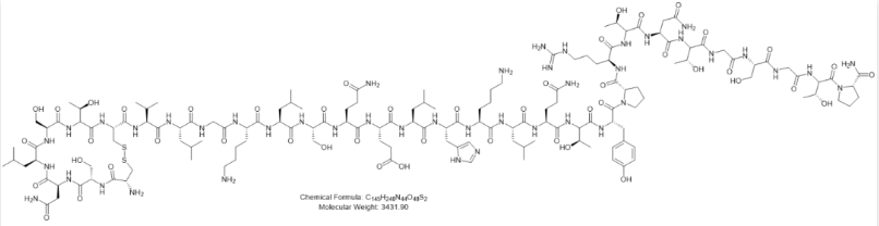
The active ingredient in Miacalcin® is the peptide calcitonin salmon (salmon calcitonin), a calcitonin hormone found in salmon. Like human calcitonin, calcitonin salmon is a peptide hormone that is secreted by the parafollicular cells of the thyroid gland in response to hypercalcemia and reduces blood calcium and phosphorus by promoting renal excretion. Miacalcin® nasal spray is used to treat osteoporosis in women who have been menopausal for at least 5 years. It should work in conjunction with adequate calcium and vitamin D intake. Miacalcin® nasal spray is aimed at patients who are not candidates for alternative therapies, such as those for whom other therapies are unsuitable or those who are intolerant or unwilling to use other therapies.
Desmopresin (trade name Stimate®)
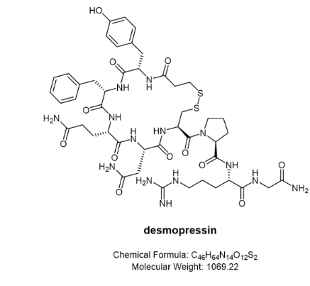
Stimate® Nasal Spray is indicated for patients with haemophilia with coagulation factor VIII activity levels greater than 5% (mild). Stimate® Nasal Spray contains desmopressin acetate, a synthetic analog of the natural hormone Arginine vasopressin, as the active substance. A single (0.1 mL, 150 μg) spray of Stimate® nasal spray solution has an anti-diuretic activity of approximately 600 IU. The half-life of Stimate® Nasal Spray is between 3.3 and 3.5 hours and the intranasal dose ranges from 150 to 450 μg. The plasma concentration of Stimate® Nasal Spray reaches a maximum of approximately 40 to 45 minutes after administration. When administered by the intranasal route as a 1.5 mg/mL solution, the bioavailability of Stimate® nasal spray ranged from 3.3% to 4.1%.
Desmopressin (trade name Octostim®)
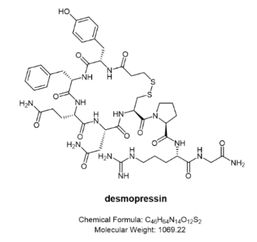
Also desmopressin, its original developer Ferring Pharmaceuticals developed it as a different nasal spray. Octostim® is an anti-diuretic, it can be used to treat central insipidus, a condition in which the kidneys are unable to retain water due to a deficiency of antidiuretic hormone (ADH). This condition causes frequent urination and thirst. It is also used to treat temporary thirst and increased urination that may occur after a head injury or certain types of brain surgery. For these uses, Desmopressin Nasal Solution works by helping to reduce the amount of urine produced by the kidneys.
Desmopressin (trade name Minirin®)
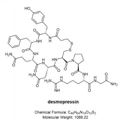
Primary nocturnal enuresis: Minirin® nasal spray is indicated for the management of primary nocturnal enuresis. It can be used alone or as an adjunct to other non-pharmacological interventions for behavioral modification. It has been shown to be effective in certain cases that are difficult to treat with conventional therapy.
Central intracranial insipidus: Minirin® nasal spray is indicated for the treatment of central intracranial diabetes insipidus and for anti-diuretic replacement therapy for temporary polyuria following head trauma or surgery in the pituitary region. It is not effective in treating nephrogenic diabetes insipidus.
The Minirin® nasal spray compression pump delivers 0.1 mL (10 μg) of desmopressin acetate per spray. In addition to the nasal spray, Minirin® is also available in the form of an oral tablet.
Desmopressin (trade name DDAVP)
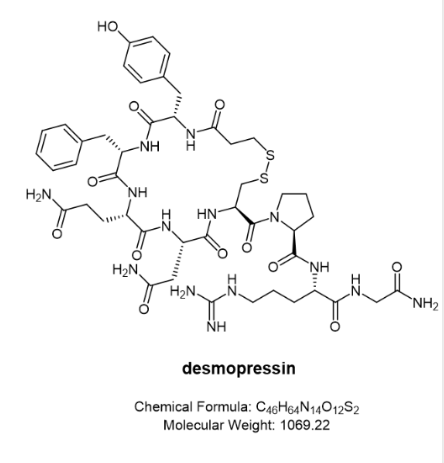
DDAVP contains desmopressin acetate as the active substance with an anti-diuretic effect and 1 mL (0.1 mg) of intranasal DDAVP is active at approximately 400 IU. The biphasic half-lives of intranasal DDAVP were 7.8 minutes and 75.5 minutes in the fast and slow phases respectively, compared to 2.5 and 14.5 minutes for the other form of lysine vasopressin. Thus, intranasal DDAVP produces a rapid antidiuretic effect and a long duration of action after each dose.
Buserelin (trade name Suprefact®)
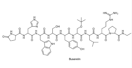
Buserelin is used in men to treat advanced prostate cancer but is not a cure. Most types of prostate cancer require the male hormone testosterone to grow and spread. Buserelin works by reducing the amount of testosterone produced by the body. This action helps to slow or stop the growth of cancer cells and helps to relieve symptoms such as painful/difficult urination.
Buserelin is also used to treat endometriosis in women. It works by reducing the body’s production of the female hormone estrogen. This effect helps to shrink abnormal tissue and reduce the symptoms of endometriosis (e.g. pelvic pain, dysmenorrhoea).
Buserelin is an artificial hormone, similar to the natural hormone “gonadotropin-releasing hormone” (GnRH) produced by the human body. Although the use of buserelin promotes the secretion of luteinizing hormone (LH) and follicle-stimulating hormone (FSH) (agonists), however, with prolonged administration of buserelin, the GnRH receptor becomes desensitized and completely stops responding to buserelin and endogenous GnRH. The profound desensitization of the GnRH receptor leads to a reduction in LH and FSH secretion from the anterior pituitary gland, followed by a cessation of gonadal sex hormone production, a significant reduction or loss of spermatogenesis in men, and an absence of ovulation in women, and the goal of “starving” cancer cells.
Suprefact contains 1.06 mg buserelin acetate (equivalent to 1 mg buserelin free base) per mL of the intranasal aqueous solution. For prostate cancer, the usual starting dose is 500 µg buserelin, by subcutaneous injection every 8 hours for 7 days. This is followed by maintenance therapy with a single daily injection of 200 µg buserelin or 400 µg of nasal spray delivered three times daily (2 sprays in each nostril). Women treated with buserelin for endometriosis are usually treated with buserelin nasal solution at a dose of 400 µg (2 sprays per nostril) 3 times a day. Treatment usually lasts for 6 months, but should not last longer than 9 months.
Nafarelin (trade name Synarel®)
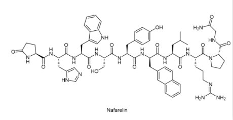
Like buserelin, nafarelin is a GnRH (gonadotropin-releasing hormone) analog (agonist). It is used to treat endometriosis and precocious puberty. It is also used to treat uterine fibroids, to control ovarian stimulation in in vitro fertilization (IVF), and as part of cross-sex hormone therapy.
Synarel® is indicated for the treatment of central precocious puberty (CPP) (gonadotropin-dependent precocious puberty) in both male and female children. It is used as a nasal spray 2 to 3 times a day. Synarel® nasal solution contains nafarelin acetate (2 mg/mL). After filling the Synarel® pump, each start of the unit delivers a spray of approximately 100 μL containing approximately 200 μg nafarelin free base. The contents of a spray bottle are provided to deliver at least 60 sprays.
After intranasal administration, nafarelin acetate is rapidly absorbed into the systemic circulation. The maximum serum concentrations were reached between 10 and 40 minutes. The average peak concentration was 0.6 ng/mL (range 0.2 to 1.4 ng/mL), after a single dose of 200 μg free base nafarelin and after a single dose of 400 μg free base nafarelin, the mean peak concentration observed was 1.8 ng/mL (range 0.5 to 5.3 ng/mL). The bioavailability of the 400 μg dose averaged 2.8% (range 1.2 to 5.6%). The mean serum half-life of nafarelin after intranasal administration is about 3 hours. Approximately 80% of nafarelin acetate is bound to plasma proteins at 4°C.[3]
Oxytocin (trade name Syntocinon)
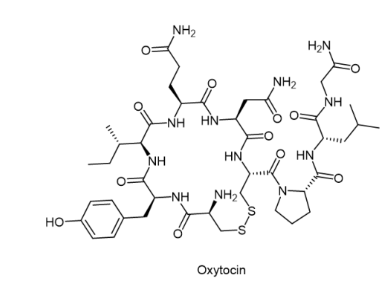
The uses of oxytocin nasal spray range from treating anxiety disorders to autism. In clinical trials, oxytocin has shown promise in the treatment of obesity and may have other super-indicated uses. It is a naturally occurring hormone that can also be used as a supplement.
Semax
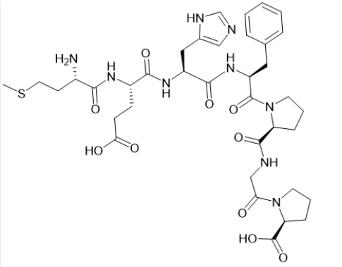
Semax peptides are known for their neuroprotective, educational, and neurorestorative properties. Traditionally, Semax has been used to treat depression and anxiety. Semax was originally developed based on the molecular structure of ACTH (Adrenocorticotropic Hormone). Ongoing research suggests that Semax may be useful for individuals with cognitive disorders (including dementia, stroke, ADHD, etc.) and may better improve mental clarity and concentration, reduce anxiety and depressive symptoms, improve the clarity of thought and focus, and reduce the effects of stress, hence Semax is given the free translation of “brain peptide” in Chinese. “Semax is used for a wide range of indications, and in the medical setting, Semax has been prescribed for the treatment of anxiety disorders, ischaemic events, stroke, neuroregeneration, ADHD, opioid withdrawal, Parkinson’s disease, Alzheimer’s disease, thrombosis, and gastric disorders. It is worth noting that Cerebrolysin is mainly used in countries such as Russia and Ukraine and is not approved in most countries around the world.
Advantages of intranasal delivery of peptide drugs
Potential nose-to-brain route: The blood-brain barrier (BBB) is a major barrier to the delivery of therapeutic agents to the central nervous system (CNS), as most large molecular weight substances are severely restricted from crossing the BBB under normal circumstances. For biological agents, such as peptides, proteins, monoclonal antibodies, oligonucleotides, and gene and cell therapies via the nasal-brain pathway, nasal delivery offers a potential strategy to bypass the BBB and reach the brain, thereby providing new therapeutic approaches for indications such as Alzheimer’s disease, Parkinson’s disease, and antipsychotic drug-induced symptoms.
High efficacy and fast onset: Most biologicals are readily degraded enzymatically in the gastrointestinal tract, therefore the most common route of administration is by injection. However, when drugs are administered by the intranasal route, they enter through the respiratory zone around the inferior turbinate, where the nasal mucosa in the respiratory zone is richly vascularised and lined with columnar epithelial cells with a large surface area (> 150 cm2 ), which is conducive to drug absorption and has strong permeability. [5] As the intranasal route avoids enzymatic degradation in the gastrointestinal tract and first-pass metabolism in the liver, as a result, it reduces the common barriers that limit drug absorption and facilitates the rapid onset of action.
High patient dependence: Intranasal delivery will undoubtedly result in higher patient compliance compared to injection. Good patient compliance is important for therapeutic outcomes, especially for intranasal drug products, which must be administered regularly and consistently to ensure sustained therapeutic outcomes. Patients’ satisfaction and comfort with the administration of medication is highly dependent on their compliance. The intranasal route is both non-invasive and highly tolerable, expanding the possibilities and compliance of patient self-administration
References
- Fosgerau, K. et al. Peptide therapeutics: current status and future directions. Drug Discov. Today. 2015, 20, 122-128.
- Arora, P. et al. Permeability issues in nasal drug delivery. Drug Discov. Today. 2002, 7, 967-975.
- Darshana, S. et al. Nasal Delivery of Proteins and Peptides. Glob. J. Pharm. Sci. 2017, 1, 555569.
- Grassin-Delyle, S. et al. Intranasal drug delivery: an efficient and non-invasive route for systemic administration: focus on opioids. Pharmacol. Ther. 2012, 134, 366-379.
- Ward, D. Optimising Nasal Drug Products for Systemic Delivery.
- Bakri, W. A., et al. Overview of intranasally delivered peptides: key considerations for pharmaceutical development. Expert Opinion on Drug Delivery. 2018, 15, 991-1005.
- Basu, S. et al. Preparation and characterization of mucoadhesive nasal gel of venlafaxine hydrochloride for treatment of anxiety disorders. Indian J Pharm Sci. 2012, 74, 428-433.
Recently, FDA approved the dual immunotherapy treatment for hepatocellular carcinoma with Astrazeneca’s Imjudo (tremelimumab) combined with Imfinzi (durvalumab), marking the second time of this year for the authoritative approval of dual immunotherapy for the treatment of cancer. The first dual immunotherapy treatment for cancer came on March 18, when FDA approved Bristol-Myers Squibb’s OpdualagTM (nivolumab and relatlimab-rmbw) to treat unresectable or metastatic melanoma.
Antibody is undoubtedly the main force in the treatment of tumors by immunotherapy including double immunotherapy. However, antibody has the disadvantages of hig cost and poor oral applicability. Fortunately, peptide-based drugs with much smaller molecular weight than antibodies could overcome the limitations and play a significant role in immunotherapy against tumors.
Peptides have a small size, high affinity, and low immunogenicity, and are easy to modify. Some modified peptides showed excellent stability, such as the antibody proteins in the two dual immunomodulators above. Peptide molecules can act as immune checkpoint inhibitors in tumor immunotherapy. Instead of attacking tumor cells directly, immunotherapy drugs modulate the patient’s immune system and attack tumor cells by targeting immune checkpoints.
What is immune checkpoint?
When proteins on the surface of T cells recognize and bind to companion proteins on other cells, immune checkpoints work. The proteins on these T cells are called immune checkpoints. When the checkpoints bind to the cancer cell’s chaperone proteins, they will send an “off” signal to the T cell. Immune checkpoint molecules matter a lot in the immune escape of tumors and can be used to restore the anti-tumor function of immune cells by blocking the binding of immune checkpoints and ligands in the clinic, so as to achieve the purpose of tumor treatment.
Since immune checkpoints can prevent overactive immune responses and sometimes hinder T cells from killing cancer cells, when these checkpoints are blocked or inhibited, T cells are better able to kill cancer cells. This result can bring a very positive effect on tumor therapy. Therefore, immune checkpoints can act as targets for cancer immunotherapy because they have the potential to be used in the treatment of many types of cancer.
Examples of checkpoint inhibitors
Currently approved checkpoint inhibitors are used to block CTLA4, PD-1, and PD-L1.
- CTLA4 inhibitors
CTLA-4 or CTLA4 (Cytotoxic T lymphocyte antigen-4), also known as CD152, is a protein receptor that acts as an immune checkpoint and downregulates immune responses. CTLA-4 is expressed in regulatory T cells but only upregulated in conventional T cells after activation. This phenomenon is particularly evident in cancer.
mAb antibody inhibitors that target the immune checkpoint CLTA4 include ipilimumab for melanoma. Many active peptides also showed a good inhibitory effect on CTLA4. For example, yeast-displayed peptide ERY2-4 specifically bound to CTLA-4 with a KD of 196.8 ± 2.3 nM. Furthermore, ERY2-4 blocked the interaction between CTLA-4 and dendritic cells (DCs) presenting B7 on their surface. Importantly, ERY2-4 showed no cross-reactivity against CD28, suggesting it does not suppress T-cell activation. Since CTLA-4 is a critical immune checkpoint for restricting the cancer immune response, this inhibitory peptide represents a new class of drug candidates for immunotherapy.
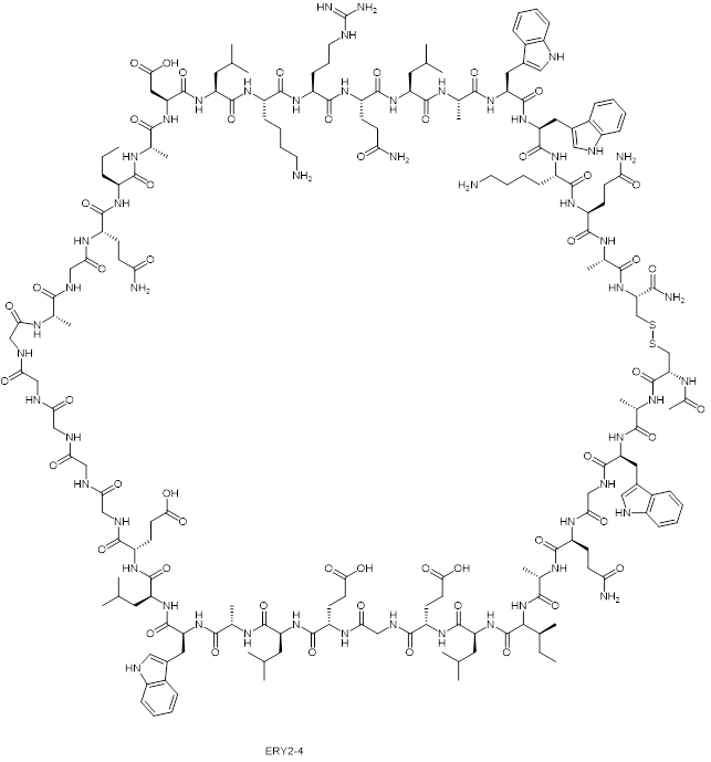
Fig1. The structure of yeast-displayed peptide ERY2-4
- PD-1
Although PD-1 and CTLA-4 belong to the CD-28 immune checkpoint receptor family, their roles in suppressing the immune response are mechanistically different. CTLA-4 regulates immune response mainly in lymph nodes in the early stage, while PD-1 regulates immunosuppression in peripheral tissues in the later stage. Nevertheless, clinically approved antibodies against both pathways produced long-lasting clinical responses, they both resulted in adverse consequences and anti-CTLA-4 antibodies showed a higher incidence and level of immune-related adverse reactions. The better clinical profile of PD-1/PD-L1 checkpoint inhibitors in terms of efficacy and safety has led to rapid drug development and approval in multiple indications. In this area, Bristol-Myers Squibb has patented their cyclic peptide, which has been shown to have nanomolar activity in inhibiting PD-1/PD-L1 interactions.
- BTLA-4
B and T lymphocyte attenuation factor (BTLA) is another co-inhibitory receptor in the CD-28 family. In a recent report, based on the natural protein HVEM (herpesvirus entry mediator), which binds to BTLA, researchers designed a disulfide bond bridging peptide using the HVEM (14-39) fragment. BTLA expressing 293T cells were used to demonstrate their cellular activity. HVEM (14 — 39) peptide is a BTLA/HVEM binding inhibitor that inhibits the formation of BTLA/HVEM complex in a dose-dependent manner. It deserves further development in the field of immunotherapy against tumors.
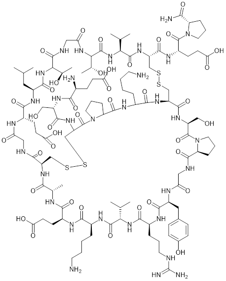
Fig2. The structure of HVEM (14-39) peptide
- VISTA
VISTA is an immune checkpoint in the B7 family that collaborates with the PD-1 protein to regulate T-cell responses. CTLA-4 and PD-1 inhibit immune function during T-cell initiation and effect, while VISTA inhibits immune response during the earliest stages of T-cell activation. To this end, the researchers designed VISTA peptide antagonists. This is a cyclic peptide containing a disulfide bond and multiple D-amino acid residues.
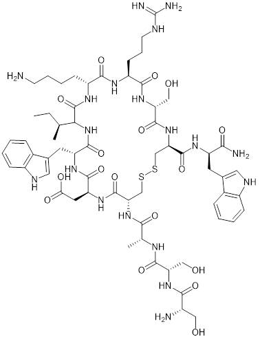
Fig3. VISTA peptide inhibitor
The peptide VISTA antagonist significantly enhanced T cell proliferation compared with anti-VISTA antibody or anti-PD-L1 antibody. In addition, the antitumor efficacy of this VISTA antagonist peptide was demonstrated in a model of immunogenic bladder cancer MB49. The founding suggests that the mechanism of action may be that peptides bind to key/active sites in VISTA, thereby blocking VISTA’s immunosuppressive function.
- LAG-3
LAG-3 is the third clinically targeted immune checkpoint receptor after CTLA-4 and PD-1. LAG-3 is co-expressed with other receptors, especially PD-1, and inhibits immune function in a variety of settings, making it an attractive target for combination with PD-1. The anti-LAG-3 and anti-PD-1 antibodies have been a success in the treatment of melanoma. Bristol Myers Squibb’s OpdualagTM, whose active ingredients nivolumab and relatliab-RMBW are antagonists of PD-1 and LAG-3, respectively, was approved by the FDA on March 18, 2022. OpdualagTM thus became the first immunotherapy targeting LAG-3 approved by the FDA.
In the study of peptide LAG-3 antagonists, researchers designed a disulfide cyclic peptide named C25, which has a KD value of mM and can effectively bind to LAG-3, thus efficiently interfere with the binding between LAG-3 and MHC-II (major histocompatibility complex protein II).
Conclusion
Immune checkpoint antagonists are currently dominated by antibodies, but low response rates and immune-related adverse consequences require the development of more effective and safer therapeutic modalities. Peptide drugs have potential advantages in selectivity, efficacy, and reduction of off-target toxicity. In the future, peptide immunotherapy may lead to the development of parallel antibody drugs, and perhaps peptide dual immunotherapy similar to OpdualagTM.
Related Products:
| Product | Description |
| Checkpoint Inhibitors | Checkpoint inhibitors are also described as a monoclonal antibody or targeted therapy. |
| Therapeutic Peptides | Therapeutic peptides are a unique class of pharmaceutical agents composed of a series of well-ordered amino acids. |
| Peptide Inhibitors | Peptide inhibitors have the advantages of high affinity and specificity, low production cost, etc., and have attracted the attention of researchers. |
Related Services:
| Service | Description |
| Pharmaceutical Peptides Services | The pharmaceutical peptides services of Creative Peptides can help you significantly shorten the development time of peptide drugs in the development of peptide drugs. |
References
- Tharanga, M. R. et al. An Immune-Stimulatory Helix–Loop–Helix Peptide: Selective Inhibition of CTLA-4–B7 Interaction. ACS Chem. Biol. 2020,15, 360–368.
- Callahan, M. K. At the bedside: CTLA-4- and PD-1-blocking antibodies in cancer immunotherapy. J Leukoc Biol, 2013, 94, 41-53.
- Liu, J. et al. Immune-checkpoint proteins VISTA and PD-1 nonredundantly regulate murine T-cell responses. Proc. Natl. Acad. Sci. U.S.A. 2015, 112, 6682-6687.
Tab 1. FDA-approved covalent peptide drugs from 2011 to 2019
| Name | Therapeutic Field | Active Gene | Chemical Structure |
| Telaprevir
|
Anti-hepatitis C virus | α-ketoamide | 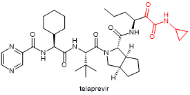 |
| Boceprevir
|
Anti-hepatitis C virus | α-ketoamide | 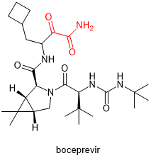 |
| Narlaprevir
|
Anti-hepatitis C virus | α-ketoamide | 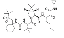 |
Covalent peptide drugs usually have electrophilic groups (because the reactive groups on the corresponding protein receptor are generally nucleophilic groups), and the reaction between the electrophilic group and the nucleophilic group of the receptor leads to covalent binding between the drug molecule and the receptor, thus achieving the therapeutic effect. The invention of covalent peptide drugs has led to the re-emergence of many protein receptors previously considered “undruggable”.
One of the advantages of covalent peptide drug development is that it can combine molecules or groups based on the previous results of non-covalent drugs and introduce active groups into the lead of non-covalently bonded peptides that bind closely to the target protein. When non-covalent binding occurs, the active groups are introduced into the vicinity of the target amino acid residue of the target protein in the three-dimensional space. In this case, the amino acid residue reacts irreversibly with the active groups on the drug molecule to achieve the ultimate goal of inhibiting the target protein (Fig 1).

Fig. 1. The covalent binding of a peptide drug to a target protein based on non-covalent interaction
From a protein receptor point of view, cysteine is the most studied target for drug designers because its sulfhydryl group acts as an excellent nucleophile. In the process of drug design, acrylamide groups are often attached to the lysine residues in the original non-covalent peptide drugs, and covalent bonds are formed by the reaction between the sulfhydryl group of the receptor cysteine and the acrylamide of the drug molecules. A study showed that a modified peptide drug (a 13-peptide containing acrylamide lysine) was more effective at inhibiting Siah (E3 ubiquitin-protein ligase) and that the peptide drug was specifically bound to cysteines at key sites. However, when the acrylamide group is replaced by the more active chloroacetamide group, the resulting peptide drug loses its cysteine selectivity and can instead react with other cysteines on the Siah protein receptor. This result confirmed that the reactivity of the electrophilic groups of covalent drugs is very important for drug selectivity.
Since cysteine is scarce in native protein receptors, covalent peptide drugs targeting lysine and histidine have gained attention. In one study, a peptide drug with a benzoyl fluoride group formed a strong covalent binding to its receptor p53-MDM2/4 protein, effectively inhibiting the activity of this protein. The activity of the modified peptide was 5-fold enhanced compared with that of the peptide precursor without benzenesulfonyl fluoride. Notably, the SuFEx reaction between benzenesulfonyl fluoride and the amino group of the lysine side chain is considered a new type of click reaction.
Encouraged by the sulfur-fluorine exchange reactions, different active groups of benzenesulfonyl fluoride and fluorosulfonic acid aromatic esters were of interest. They can covalently bind to lysine, tyrosine, and histidine.
In the search for drugs to treat COVID-19, covalent peptide drugs have also emerged. A peptide molecule named VIR251, containing methyl acrylate structure, can covalently interact with papain-like protease (PLpro) of SARS-CoV-2 protein mediating COVID-19 through Michael’s addition. Thus, the physiological function of the protein was inhibited.
Conclusion
With the continuous development of small molecule covalent drugs and the R&D of non-covalent peptide drugs, the resources available for the combination of the two products, covalent peptide drugs, will continue to expand, and promote the R&D and marketing of new drugs in this field. However, while covalently bonded drugs have the advantage of long duration and low dose, practitioners are still concerned about the unanticipated side effects of these active substances indiscriminately binding to proteins in the body, as well as the possible immune response to these drugs. Therefore, safety has become a top priority in the design and development of covalently bonded drugs.
Related Products:
| Name | Description |
| Therapeutic Peptides | Creative Peptides is the world’s first-choice supplier of peptides used as pharmaceutical agents with a range of more than 100 peptides and related products. |
| Cysteine | Cysteine is a semiessential proteinogenic amino acid with the formula HOOC-CH-(NH2)-CH2-SH. |
| Lysine | Lysine is an α-amino acid that is a precursor to many proteins. |
| Histidine | Histidine is an essential α-amino acid that is used in the biosynthesis of proteins. |
| Tyrosine | Tyrosine is a type of amino acid, which are the building blocks of protein. |
References
[1] Dong J, Krasnova L, Finn MG, et al. Sulfur (VI) Fluoride Exchange (SuFEx): Another Good Reaction for Click Chemistry. Angewandte Chemie Int Ed. 2014, 53,9430-9448.
[2] Hoppmann C, Wang L. Proximity-enabled bioreactivity to generate covalent peptide inhibitors of p53-Mdm4. Chem. Commun. 2016, 52, 5140-5143.
[3] Pietro Delre, Fabiana Caporuscio, Michele Saviano, Giuseppe Felice Mangiatordi, Repurposing Known Drugs as Covalent and Non-covalent Inhibitors of the SARS-CoV-2 Papain-Like Protease. J. Med. Chem. 2019, 62, 9188-9200.
[4] Stebbins JL, Santelli E, Feng Y, et al. Structure-Based Design of Covalent Siah Inhibitors. Chem Biol. 2013, 20, 973-982.
[5] Wang N, Yang B, Fu C, et al. Genetically Encoding Fluorosulfate-l-tyrosine To React with Lysine, Histidine, and Tyrosine via SuFEx in Proteins in Vivo. J. Am. Chem. Soc. 2018, 140, 4995-4999.
]]>- Peptide drugs for cancer immunotherapy
The treatment of cancer by controlling the function of immune cells is a research hotspot of peptide drugs. PD-L1 is typically overexpressed on many cancer cells and when it binds to PD-1 expressing on activated T cells, the cancer cells can escape from the immune response. Therefore, inhibition of PD-1/PD-L1 interaction could be a method of tumor immunotherapy. Studies have shown that hydrophilic D-type peptide (D-PPA) itself has no cytotoxicity. When blocking the PD-1/PD-L1 interaction, it can inhibit tumor growth in CT26 tumor-bearing mice and prolong their lifespan.
Macrophages are immune cells that play an important role in innate and acquired immunity. The CSF-1/CSF-1R pathway is related to the survival and differentiation of macrophages. In a recent study, a peptide called M2pep[Cys-YeqDPwGVKWWY] was modified to improve specificity against M2 TAMs. The experimental results confirmed that co-encapsulation with CSF-1R siRNA and M2pep could target M2 TAM to remodel into the M1 phenotype and increase anti-tumor immune response.
- Apoptosis-related peptide drugs
Certain peptides can induce apoptosis in cancer cells. For example, an anticancer peptide called RA-V (deoxybouvardin) can induce mitochondrial apoptosis in tumor cells, followed by loss of mitochondrial function. Another pentapeptide, Dolastatin 10, was able to induce apoptosis by up-regulating cytochrome C and Bax.
KLA (KLAKLAKLAK) is also one of the pro-apoptotic peptides, which induces programmed cell death by disrupting the mitochondrial membrane. Bioconjugates containing KLA peptides can be used in the development of various anticancer drugs. For example, KLA can bind to the tumor homing peptide IRGD(CRGDKGPDC) to improve the permeability of tumor tissues and cells. The potent anti-tumor effect of the recombinant KLA-IRGd conjugate as well as the high tumor selectivity and low systemic toxicity have been demonstrated in mouse models. These studies may lead to the development of novel targeted PDCS.
Another example of a KLA-based therapy is the penetratin (a cell-penetrating peptide)-KLA conjugate. To overcome the low cellular permeability of KLA peptides, penetratin was linked to KLA peptides via disulfide bonds, resulting in high cellular permeability and cytotoxicity, even at low concentrations. The penetratin-KLA conjugate has little effect on the mitochondria of normal cells, showing its therapeutic potential as a novel peptide conjugate.
- Peptides for tumor enrichment
There have also been some new studies describing various methods of targeting tumors with peptides. For example, it was reported that the S-S-bridged cyclic peptide (ALOS-4) is specific for the integrin AVβ3, which is highly expressed in human metastatic melanoma. In this study, ALOS-4 was coupled to the topoisomerase I inhibitor camptothecin. The results showed that the conjugate had potent anti-tumor activity in human metastatic melanoma cells, but low toxicity to normal cells. In addition, a researcher team conjugated the four-branched peptide NT4 to paclitaxel (PTX). NT4 peptides can selectively bind to tumor membrane glycosaminoglycans sulfate. It is highly selective for cancer cells when combined with paclitaxel, and this combination allows it to exhibit more potent anti-tumor activity than paclitaxel alone.
In another study, the tumor targeting of albumin-binding peptides was investigated. In vitro studies, the bioconjugation of DICLPRWGCLW to form a stable albumin complex can play a role in tumor targeting, and also showed high tumor targeting and significantly increased half-life in the SCC7 tumor-bearing mouse model. Based on the positive correlation between albumin uptake and tumor growth, albumin binding is expected to become a novel therapeutic strategy for targeting tumor tissues.
- Peptide drugs conjugated with small cytotoxic drugs
Under the concept of drug conjugates, the combination of peptides with cytotoxic drugs or other therapeutic agents can be used to develop peptide-based therapeutic agents.
Several studies have shown that cytotoxic drugs can bind to peptides with additional functions. For example, in anticancer therapy, platinum-based cisplatin chemotherapy is one of the most widely used cancer treatment strategies. However, because of cisplatin resistance, long-term use of cisplatin in patients reduces its efficacy. The use of peptides may be an important way to solve the problems related to drug resistance. When cisplatin is combined with peptides, the binding induces a high accumulation of cisplatin in tumors and metastasis-containing organs and significantly reduces the systemic toxicity of cisplatin.
Several peptide drug conjugations are currently under clinical investigation for the treatment of refractory tumors. On February 27, 2021, Melflufen, the first peptide-coupled drug, was approved by FDA for the treatment of multiple myeloma. Because melflufen is highly lipophilic, it is readily ingested by targeted cancer cells. Once internalized, melflufen molecules can be cleaved by aminopeptidase, which is highly expressed in multiple myeloma cells, and release alkylating agent toxic drugs that cause DNA damage and apoptosis in tumor cells. Now, many other forms of peptide-drug conjugates are also under active development as anticancer drugs.
Creative Peptides’ Perspectives
Due to the advantages of peptides, peptide drugs or peptide-drug conjugates have been extensively explored for new drug R&D. Previously, peptide drugs were seen as simply mimicking hormones or endogenous peptides in the body, but they are now being reborn to be designed into new therapies. However, as the limitations of peptide drugs have been solved, the powerful anti-tumor effect of peptide drugs has attracted the attention of researchers and the pipeline layout of more and more companies. Various therapeutic strategies that combine cytotoxic antitumor drugs with functional peptides come to a reality and are expected to be more effective than small-molecule drugs, antibodies, and ADCs.
References:
- Blocking of the PD-1/PD-L1 Interaction by ad-Peptide Antagonist for Cancer Immunotherapy.
- Tumor-penetrating peptide fused to a pro-apoptotic peptide facilitates effective gastric cancer therapy.
Related Products:
| Name | Description |
| Cell Penetrating Peptides | Most CPPs are relatively short peptides that consist of less than 40 amino acids and are able to enter cells through various ways. |
| Anti-Tumor Peptides | The potential use of peptides as anticancer drugs are highlighting current scenario and future prospects. |
| Dolastatin 10 | A pentapeptide originally isolated from the marine mollusk Dolabella auricularia with potential antineoplastic activity. |
| iRGD peptide | iRGD peptide is a 9-amino acid cyclic peptide, and has tumor-targeting and tumor-penetrating properties. |
| Penetratin | Penetratin is a cell-penetrating peptide (CPP), also known as a protein transduction domain (PTD). |