Basic Principles of SPPS
Peptide synthesis involves a large number of repetitive steps, and the use of a solid support offers significant advantages. In such a system, high concentrations of excess reagents can drive coupling reactions to completion, while surplus reagents and by-products can be separated from the insoluble growing peptide chain (resin) simply by filtration and washing. All operations are carried out in the same solvent, eliminating the need for material transfer.
- Anchoring of the initial amino acid: The initial amino acid is anchored to the resin through its C-terminus, forming either an ester bond or an amide bond.
- Linear assembly: The peptide chain is elongated stepwise from the C-terminus to the N-terminus through condensation reactions.
- Protecting-group strategy: Permanent protecting groups are used for side chains to improve stability; non-side-chain amino acid protection is generally achieved using carbamate-type protecting groups, which can be removed under mild conditions, thereby avoiding racemization caused by overly harsh conditions.
- Cyclic operations: Coupling, washing to remove excess reagents, deprotection, and initiation of the next coupling cycle.
- Final cleavage: The peptide chain is cleaved from the resin while simultaneously removing the side-chain protecting groups.
Fmoc/t-Bu Strategy in Solid-Phase Synthesis
Two major protecting-group strategies are mainly used in solid-phase synthesis: Boc/Bzl and Fmoc/t-Bu. The former requires deprotection under relatively strong acidic conditions, such as TFA, and is mainly used for side-chain protection. Strong acidic conditions often damage sensitive amino acid sequences; therefore, the Fmoc/t-Bu strategy is generally adopted for amino acid protection, in which Fmoc is base-labile and t-Bu is weak-acid-labile. Solid-phase synthesis methods using the Fmoc-based strategy have become the preferred approach for routine peptide synthesis.
Solid Supports
Solid supports can be characterized by both the “matrix polymer” and the “linker molecule,” although the term “resin” is often incorrectly used to refer to the entire system. In solid-phase chemistry, the matrix polymer is as important as the liquid-phase environment. At present, there are well over one hundred commercially available resins, some of which are equipped with the same linker molecules. Therefore, the most suitable linker must be selected carefully during synthesis.
Cross-linked polystyrene (PS)-based resins are commonly used solid supports for SPPS. Resin beads with a particle size distribution of 200-400 mesh (approximately 50 μm in diameter) and a loading of 0.5-0.8 mmol/g exhibit good swelling properties in DMF and DCM, which promotes diffusion of reactants into the polymer matrix and ensures adequate access to linker sites buried within the beads. For longer peptide chains (more than 25 amino acids) or sequences that are more difficult to synthesize, lower-loading resins of 0.1-0.2 mmol/g should be used. Cross-linked polyamide (PA)-based resins and polystyrene-polyethylene glycol (PS-PEG) composite resins are more hydrophilic solid supports. Their microscopic and macroscopic physical properties differ from those of PS resins. These supports generally have lower loading capacities and may serve as alternatives to PS for the synthesis of complex sequences and large peptides.
Resin Handling
Why must resins be treated before use? This is analogous to textile dyeing and finishing. Because cotton fibers have irregular oval cross-sections, they must be swollen before dyeing to increase the surface area so that dyes can penetrate effectively. Resin is a polymeric material with long chains that may become entangled or structurally collapsed, and its reactive sites are located inside the resin. Therefore, swelling treatment before use is essential, and the reaction conditions must also be taken into consideration.
Selection of the Reactor
SPPS reaction vessel: The reactor is generally made of glass and equipped with a PTFE or glass frit syringe. The size of the reactor should be selected according to the amount of resin used.
| Container Length (cm) | Container Diameter (cm) | Maximum Resin Weight (g) | Working Volume (mL) |
| 5 | 2 | 0.5 | 10 |
| 11 | 2.6 | 2 | 40 |
| 15 | 3.4 | 4 | 90 |
SPPS Reactor Types
Selection of Solvents
Ninety-nine percent of the coupling sites are not located on the surface of the resin beads but rather inside the resin. Therefore, resin beads bearing the growing peptide chain must be fully swollen to ensure optimal penetration of the protected activated amino acids into the polymer matrix, thereby improving coupling efficiency.
The basic swelling procedure is as follows: before starting solid-phase synthesis, place the resin in a suitable solvent such as DMF or DCM and allow it to swell for 20-30 minutes. For cross-linked polystyrene (PS)-based resin beads, DCM provides the best swelling characteristics. During the coupling step, however, DMF or NMP is preferred because these solvents provide better solubility for the reactants. Alcohols and water are not suitable for cross-linked polystyrene (PS)-based resins. However, in subsequent washing steps, methanol or isopropanol may be used to shrink the resin beads and expel entrapped excess reactants. After this treatment, the beads must be reswollen with DCM and DMF. Resin swelling procedure: Place the resin in a suitable reactor, add DCM until the resin beads are fully immersed, and gently mix the resin suspension with a PTFE stirring rod. Allow it to swell for 20-30 minutes, then remove the solvent by vacuum filtration.
Stirring and Mixing
During SPPS, reaction kinetics are governed primarily by diffusion phenomena; therefore, vigorous stirring of the reaction vessel is unnecessary. In addition, most resin beads used in peptide synthesis are relatively fragile, and strong agitation can damage or fracture them. Gentle mixing may be achieved using the rotor of a rotary evaporator or by employing devices capable of mild motion such as rocking or vortex-like mixing.
Washing
Washing is used to remove soluble by-products and excess reagents generated during coupling and deprotection. The basic washing procedure involves introducing solvent into the reactor and then removing it under vacuum; when necessary, the resin may be stirred gently in the washing solvent with a stirring rod.
Standard washing and reswelling procedure: Add DMF to the reaction vessel, allow it to stand for 10 s, then remove the solvent by vacuum filtration, carefully rinsing all parts of the reactor with DMF. Remove the solvent under vacuum, then add fresh solvent again for washing, repeating the operation 1-2 times. After washing is complete, wash the resin with methanol using a procedure similar to the DMF washing step, repeating 1-2 times. Then switch to DCM for washing 1-2 times, followed by DMF washing 1-2 times.
Linkers and Resin Selection in Fmoc Solid-Phase Synthesis
The first step in SPPS is to anchor the C-terminal amino acid residue (with its N-terminus protected) to the solid support through either an ester bond or an amide bond, depending on whether the target peptide has a C-terminal carboxylic acid or a C-terminal amide group. At present, most linker molecules are commercially available and preloaded onto different matrices, including polystyrene (PS), polyamide (PA), and polystyrene-polyethylene glycol (PS-PEG) composite matrices. C-terminal peptide acids are generated through ester linkages, whereas C-terminal peptide amides are generated through amide linkages. These preloaded linkers simplify experimental preparation and ensure batch-to-batch consistency. Different matrices have different characteristics: PS matrices are highly hydrophobic and suitable for routine peptide synthesis; PS-PEG matrices are more hydrophilic and advantageous for long-peptide synthesis; and polyamide (PA) is used for specialized applications.
Peptide Amides
For the synthesis of C-terminal peptide amides, the following resins are commonly used. The attachment of the first amino acid to these resins is basically consistent with the Fmoc synthesis strategy, and under TFA treatment these resins leave an amide bond upon cleavage to complete the release of the peptide (the resin functions similarly to a protecting group). Because these resins contain free amino groups, they are usually Fmoc-protected and must be deprotected before use. It should be noted that when amino acids with substantial steric hindrance at the C-terminus are coupled to the resin, incomplete conversion may occur, and a second coupling step may be required.
Peptide Acids
Compared with the nucleophilicity of amino groups, hydroxyl groups are less nucleophilic. Therefore, anchoring amino acids onto a solid support in the form of esters is more challenging. It is recommended to directly purchase resins preloaded with the protected C-terminal amino acid to avoid issues such as epimerization, dipeptide formation, and substitution-level variability. When synthesizing C-terminal peptide acids using the Fmoc/tBu strategy, the anchoring reaction must be carried out under anhydrous conditions, and amino acids containing moisture must be dried in advance.
Hydroxyl Resins
Among hydroxyl resins, the less sterically hindered Wang resin more readily forms ester bonds, and the symmetrical anhydride method is commonly used. If anchoring is difficult, the esterification step may be repeated with fresh reagents; some amino acid derivatives may require three esterification cycles to achieve satisfactory results. After anchoring is completed, the unreacted hydroxyl groups on the resin are recommended to be capped with benzoic anhydride or acetic anhydride. Standard operating procedure for hydroxyl resins: First place the resin in the reactor and swell it with a suitable solvent. Meanwhile, dissolve the target Fmoc-protected amino acid (10.0 eq.) in DCM at 0°C, add DIC (5.0 eq.) and allow activation for 10 minutes; DMF may be added if necessary to aid dissolution. Transfer the activated solution to the resin, add DMAP/DMF to catalyze the reaction, and stir for 1 hour; then wash once each with DMF and DCM. Dry under vacuum for 18 hours and determine the loading by the Fmoc release assay. If the loading is below 70%, the esterification step should be repeated.
Once the target loading is achieved, cap the residual hydroxyl groups with benzoic anhydride/acetic anhydride (5 eq.) and pyridine (1 eq.) in DMF for 30 minutes. After final washing and Fmoc deprotection, peptide-chain elongation may begin. This procedure ensures coupling efficiency through low-temperature activation, strict stoichiometric control, and loading verification. Key steps include activation monitoring, catalytic enhancement, and hydroxyl capping to prevent by-product formation.
Trityl-Series Resins
These resins exhibit high acid lability, and the steric hindrance of their linker groups can significantly suppress diketopiperazine (DKP) formation. They are particularly recommended for the synthesis of peptides with C-terminal proline (Pro) or glycine (Gly). These resins can be cleaved under extremely mild acidic conditions, thereby protecting both the peptide segment and the resin. Commercial products are generally supplied as chlorotrityl resin or trityl alcohol precursors.
Typical procedure for trityl resin: Place 1 g of trityl resin (loading 1.0-2.0 mmol chloride/g) into an SPPS reactor and allow it to swell. Add 3 equivalents of the Fmoc amino acid and 7.5 equivalents of DIPEA, then stir at room temperature in anhydrous DCM for 30-60 minutes. Wash the resin sequentially with DMF 2-3 times, then add 10 mL of a DCM/MeOH/DIPEA mixture to cap the residual chloride groups, and continue the reaction for 15 minutes. Finally, wash three times each with DMF and DCM, dry under vacuum, and determine the loading by the Fmoc release assay. Through this procedure, precise control of the amino acid-to-DIPEA ratio, together with methanol quenching, ensures single-site attachment. It should be noted that the activity of trityl chloride must be maintained under anhydrous conditions.
]]>Structural Features and Pharmacological Advantages
Macrocyclic peptides are a class of peptide molecules characterized by a ring-like structure, typically composed of 5-17 amino acid residues with a molecular weight ranging from 500 to 2000 Da. Positioned between small-molecule compounds and large biological therapeutics, they combine the structural and functional advantages of both, making them a promising direction in peptide drug discovery. The cyclic architecture endows these peptides with exceptional target-binding affinity and biological stability, enabling them to effectively modulate protein-protein interactions (PPIs) and intrinsically disordered proteins (IDPs)—targets traditionally considered “undruggable.” These features highlight their vast potential in the development of orally available peptide drugs.
Enhanced Bioactivity, Stability, and Selectivity
Compared with linear peptides, macrocyclic peptides demonstrate higher target specificity and biological activity, allowing for potent therapeutic effects at lower doses while minimizing off-target interactions and systemic toxicity. Their rigid cyclic structure improves resistance to gastric acid and proteolytic enzymes, prolonging in vivo half-life and enhancing pharmacodynamic persistence. Moreover, macrocyclic peptides exhibit remarkable structural diversity and design flexibility, allowing chemical modifications to meet diverse therapeutic needs. With low immunogenicity, strong target selectivity, and minimal impact on normal cells, they are emerging as safe and effective candidates for next-generation precision therapeutics. Continuous advancements in delivery technologies further enhance their clinical feasibility.
With their superior pharmacological profiles and evolving synthetic technologies, macrocyclic peptides have become one of the hottest frontiers in peptide drug research. They hold the promise of achieving precise target engagement, improved safety profiles, and optimized efficacy, positioning themselves as a cornerstone in the future of innovative therapeutic strategies.
Clinical Pipeline of Macrocyclic Peptide Drugs
The number of macrocyclic peptide drugs entering clinical development has been steadily increasing (Table 1), with orally administered macrocyclic peptides emerging as a major focus area. Since the approval of Cyclosporine A by the FDA in 1983, macrocyclic peptide therapeutics have made remarkable advances across multiple therapeutic fields. However, orally bioavailable macrocyclic peptides remain relatively rare. With the rapid emergence of new technologies, the development of oral peptide therapeutics is expected to achieve significant breakthroughs in the coming years. Several oral macrocyclic peptide candidates have now advanced into clinical trials, with representative examples including the following:
01. MK-0616 (Merck & Co.)
MK-0616 is an oral PCSK9 inhibitor currently in Phase III clinical trials. Designed as a macrocyclic peptide capable of binding to PCSK9 (Proprotein Convertase Subtilisin/Kexin Type 9), MK-0616 aims to reduce low-density lipoprotein cholesterol (LDL-C) levels. In contrast to current PCSK9-targeting antibody therapies that require subcutaneous injection, MK-0616’s oral formulation is expected to greatly improve patient adherence and reduce treatment costs. Although the peptide itself has limited permeability, the inclusion of an absorption enhancer allows an oral bioavailability of approximately 2%. On June 9, 2025, Merck announced that both pivotal Phase III trials—CORALreef HeFH and CORALreef AddOn—achieved their primary endpoints in patients with hypercholesterolemia, marking a major milestone for oral peptide drug development.
02. JNJ-2113 (Johnson & Johnson)
JNJ-2113 is an oral IL-23 receptor antagonist currently in Phase III clinical development. The program originated from a collaboration between Protagonist Therapeutics and Johnson & Johnson, established in 2017, with J&J leading subsequent development and commercialization. In November 2024, JNJ-2113 achieved positive topline results in its pivotal ICONIC-LEAD Phase III trial, demonstrating robust efficacy and oral potential in patients with moderate-to-severe plaque psoriasis (PsO), including adolescents aged 12 years and older. If successfully approved, JNJ-2113 is poised to become the first oral peptide drug targeting the IL-23 receptor, representing a transformative step in the treatment of chronic inflammatory diseases.
03. LUNA18 (Chugai Pharmaceutical)
LUNA18 is a KRAS inhibitor currently in Phase I clinical trials in Japan and the United States. In preclinical studies, LUNA18 demonstrated oral bioavailability ranging from 21% to 47% across multiple species (mice, rats, monkeys, and dogs), without the need for permeability enhancers—indicating excellent membrane penetration and drug-like properties. Moreover, dose-dependent antitumor activity was observed in animal models, underscoring its strong therapeutic potential as a next-generation macrocyclic peptide-based KRAS inhibitor.
| Drug Name | Highest Clinical Phase | Source Type | Clinical Trial ID | Route of Administration |
| AP301 | Phase III | Natural product derivative | NCT04551300 | Oral |
| BMS-986229 | Phase III | mRNA display | NCT04161781 | Injectable |
| JNJ-2113 | Phase III | Phage display | NCT05364554 | Oral |
| MK-0616 | Phase III | mRNA display | NCT05952896 | Oral |
| ORMD-0801 | Phase III | Natural product derivative | NCT04817215 | Oral |
| PL9643 | Phase III | Natural product derivative | NCT05201170 | Ophthalmic |
| Plitidepsin | Phase III | Natural product source | NCT01102426 | Injectable |
| Balixafortide | Phase III | Natural product derivative | NCT03786094 | Injectable |
| Rusfertide | Phase III | Natural product derivative | NCT04202965 | Injectable |
| BT8009 | Phase II/III | Phage display | NCT04561362 | Injectable |
| VT1021 | Phase II/III | Natural product derivative | NCT03970447 | Injectable |
| ALRN-6924 | Phase II | Stapled peptide design | NCT02264613 | Injectable |
| AMY-101 | Phase II | Phage display | NCT03694444 | Injectable |
| AZP-3813 | Phase II | mRNA display | Injectable | |
| Certepediol | Phase II | Phage display | NCT03517116 | Injectable |
| Dolcamatide | Phase II | Natural product derivative | NCT01983306 | Oral |
| PL8177 | Phase II | Natural product derivative | NCT05466890 | Oral |
| THR-149 | Phase II | Phage display | NCT04527107 | Injectable |
| TE-232 | Phase II | Natural product derivative | NCT00422786 | Injectable |
| BHV-1100 | Phase I/II | mRNA display | NCT04634435 | Injectable |
| BT1718 | Phase I/II | Phage display | NCT03486730 | Injectable |
| FOG-001 | Phase I/II | Phage display | NCT05919264 | Injectable |
| Lonodelestat | Phase I/II | NCT03748199 | Inhalation | |
| BT5528 | Phase I/II | Phage display | NCT04180371 | Injectable |
| BT7480 | Phase I/II | Phage display | NCT05163041 | Injectable |
| LUNA18 | Phase I | mRNA display | NCT05012618 | Oral |
Cyclic Peptides at Creative Peptides
| CAT# | Product Name | M.W | Molecular Formula |
| 10-101-103 | Vancomycin | 1449.25 | C66H75Cl2N9O24 |
| 10-101-104 | Teicoplanin | 1879.66 | C88H95Cl2N9O33 |
| 10-101-112 | Bremelanotide | 1025.18 | C50H68N14O10 |
| 10-101-169 | Pasireotide | 1047.20624 | C58H66N10O9 |
| 10-101-186 | Romidepsin | 540.69584 | C24H36N4O6S2 |
| 10-101-325 | Semaglutide | 4113.57 | C187H291N45O59 |
| 10-101-62 | Ziconotide | 2639.13 | C102H172N36O32S7 |
| 10-101-78 | Dalbavancin | 1816.69 | C88H100Cl2N10O28 |
| AF083 | Polymyxin B | ||
| MFP-041 | Rezafungin | 1226.4 | C63H85N8O17 |
| R04030 | Cyclo(-Arg-Gly-Asp-D-Phe-Val) | 574.64 | |
| R1574 | Octreotide | 1019.24 | C₄₉H₆₆N₁₀O₁₀S₂ |
| R1812 | Lanreotide | 1096.33 | C54H69N11O10S2 |
| R1824 | Cyclo(RGDyK) | C31H43F6N9O12 | |
| R2018 | Capreomycin | ||
| R2029 | Enviomycin | 685.69 | C26H43N11O11 |
| R2052 | Zilucoplan | (C2H4O)nC126H186N24O32 | |
| R2238 | Telavancin | 1755.6 | C80H106Cl2N11O27P |
| R2239 | Oritavancin | 1793.1 | C86H97Cl3N10O26 |
| R2240 | Bacitracin | 1422.69 | C66H103N17O16S |
Cell-penetrating peptides (CPPs) are short peptides capable of entering cells without disrupting membrane integrity. They typically consist of 5 to 30 amino acids and are rich in basic amino acids, including arginine and lysine. Under physiological conditions, CPPs share two common characteristics: a positive charge and amphiphilicity. In recent years, CPPs have been widely applied in drug delivery systems.
The recognized mechanisms by which CPPs penetrate cell membranes fall into two categories: endocytosis and direct translocation. Endocytosis is an energy-dependent process and includes three main pathways: macropinocytosis, caveolae-mediated endocytosis (CvME), and clathrin-mediated endocytosis (CME). In contrast, direct translocation is an energy-independent process facilitated by electrostatic interactions between the positively charged CPPs and the negatively charged membrane components and phospholipid bilayer, promoting direct membrane permeation. Several models have been proposed for direct penetration, including the barrel-stave model, carpet model, toroidal pore model, and inverted micelle model.
A summary of the cell-penetrating peptide used in drug delivery is as follows:
1. Polyarginine
Single-letter code: H2N-RRRRRRRRR-OH
Three-letter code: H2N-Arg-Arg-Arg-Arg-Arg-Arg-Arg-Arg-Arg-OH
Number of amino acids: 9
Molecular formula: C54H110N36O10
Average molecular weight (MW): 1423.69
2. Transportan 10 (TP10)
Single-letter code: H2N-AGYLLGKINLKALAALAKKIL-OH
Three-letter code: H2N-Ala-Gly-Tyr-Leu-Leu-Gly-Lys-Ile-Asn-Leu-Lys-Ala-Leu-Ala-Ala-Leu-Ala-Lys-Lys-Ile-Leu-OH
Number of amino acids: 21
Molecular formula: C104H184N26O24
Average molecular weight (MW): 2182.73
3. Tumor-Lineage Homing CPP
Single-letter code: H2N-RLYMRYYSPTTRRYG-OH
Three-letter code: H2N-Arg-Leu-Tyr-Met-Arg-Tyr-Tyr-Ser-Pro-Thr-Thr-Arg-Arg-Tyr-Gly-OH
Number of amino acids: 15
Molecular formula: C89H135N27O23S1
Average molecular weight (MW): 1983.26
4. dNP2
Single-letter code: H2N-CKIKKVKKKGRKKIKKVKKKGRK-OH
Three-letter code: H2N-Cys-Lys-Ile-Lys-Lys-Val-Lys-Lys-Lys-Gly-Arg-Lys-Lys-Ile-Lys-Lys-Val-Lys-Lys-Lys-Gly-Arg-Lys-OH
Number of amino acids: 23
Molecular formula: C125H245N43O24S1
Average molecular weight (MW): 2766.62
5. RVRR
Single-letter code: H2N-RVRR-OH
Three-letter code: H2N-Arg-Val-Arg-Arg-OH
Number of amino acids: 4
Molecular formula: C23H47N13O5
Average molecular weight (MW): 585.70
6. L-penetratin
Single-letter code: H2N-RQIKIWFQNRRMKWKK-OH
Three-letter code: H2N-Arg-Gln-Ile-Lys-Ile-Trp-Phe-Gln-Asn-Arg-Arg-Met-Lys-Trp-Lys-Lys-OH
Number of amino acids: 16
Molecular formula: C104H168N34O20S1
Average molecular weight (MW): 2246.73
7. Activable CPP (ACPP)
Single-letter code: H2N-CRRRRRRRRGGGPKKKKKK-OH
Three-letter code: H2N-Cys-Arg-Arg-Arg-Arg-Arg-Arg-Arg-Arg-Gly-Gly-Gly-Pro-Lys-Lys-Lys-Lys-Lys-Lys-OH
Number of amino acids: 19
Molecular formula: C98H191N49O20S1
Average molecular weight (MW): 2407.95
8. LH2
Single-letter code: Ac-LHHLCHLLHHLCHLAG-CONH2
Three-letter code: Ac-Leu-His-His-Leu-Cys-His-Leu-Leu-His-His-Leu-Cys-His-Leu-Ala-Gly-CONH2
Number of amino acids: 16
Molecular formula: C85H131O17N29S2
Average molecular weight (MW): 1895.26
9. Cancer Cell Line Specific CPP
Single-letter code: H2N-RLYMRYYSPTTRRYG-OH
Three-letter code: H2N-Arg-Leu-Tyr-Met-Arg-Tyr-Tyr-Ser-Pro-Thr-Thr-Arg-Arg-Tyr-Gly-OH
Number of amino acids: 15
Molecular formula: C89H135N27O23S1
Average molecular weight (MW): 1983.26
10. T2
Single-letter code: H2N-FKKFFRKLL-OH
Three-letter code: H2N-Phe-Lys-Lys-Phe-Phe-Arg-Lys-Leu-Leu-OH
Number of amino acids: 9
Molecular formula: C63H99N15O10
Average molecular weight (MW): 1226.55
Cell-penetrating Peptides (CPPs) at Creative Peptides
- Antennapedia Peptides
- Chimeric Peptides
- HIV-TAT Family
- Nuclear Localization Peptides
- Oligoarginine Peptides
People also like
- Cell Targeting Peptides: Definition, Classification and Application
- Cell-penetrating peptides (CPPs) for Diagnostics and Imaging
- Cell-penetrating Peptides (CPPs) for Gene Therapy
- Cell-penetrating Peptides (CPPs) for Drug Delivery
- Cyclic Cell Penetrating Peptides: Definition, Structure, Mechanism and Applications
- The Design of Cell Penetrating Peptides
- Classes and Applications of Cell-Penetrating Peptides
1. RGD
The tripeptide RGD can specifically bind to integrin receptors. Integrins are composed of two subunits (α and β). The integrin family, especially αVβ3, plays a crucial role in tumor angiogenesis and metastasis. During angiogenesis, these receptors are overexpressed on endothelial cells but are barely detectable in most normal organs. Therefore, they are widely used in diagnostic imaging.
Single-letter code: H2N-RGD-OH
Three-letter code: H2N-Arg-Gly-Asp-OH
Number of amino acids: 3
Molecular formula: C12H22N6O6
Average molecular weight (MW): 346.34
2. Bombesin (BBN) / Gastrin-Releasing Peptide (GRP)
BBN and its related peptides from amphibians form a neuropeptide family with multiple physiological effects, such as exocrine and endocrine secretion, thermoregulation, sucrose regulation, and cell growth. Bombesin-like peptide receptors have four subtypes: neuromedin B receptor, bombesin receptor subtype 3, gastrin-releasing peptide receptor (GRPR), and bombesin receptor subtype 4. These receptors are overexpressed in various tumors, including breast cancer, ovarian cancer, and gastrointestinal stromal tumors.
| CAT# | Product Name |
| B07004 | Bombesin (8-14) |
| B07010 | [Lys3]-Bombesin |
| B07011 | [Tyr4, D-Phe12]-Bombesin |
| B07015 | (D-Cys6,Asn7,D-Ala11,Cys14)-Bombesin (6-14) |
| B07016 | (D-Phe12)-Bombesin |
| B07017 | (D-Phe12,Leu14)-Bombesin |
| B07019 | (D-Phe6,Leu-NHEt13,des-Met14)-Bombesin (6-14) |
| B07020 | (D-Tyr6,betaPhe11,Phe13,Nle14)-Bombesin (6-14) (free acid) |
| B07022 | (Tyr4)-Bombesin |
| B07023 | Cyclo(-D-Phe-His-Trp-Ala-Val-Gly-His-Leu-Leu) |
| B07024 | Biotin – Bombesin |
| B07025 | Biotin – LC – LC – Bombesin |
| B07026 | Bombesin, FAM – labeled |
Gastrin-Releasing Peptide (GRP)
3. Somatostatin (SST) Peptides
Somatostatins (SSTs) are naturally occurring cyclic peptide hormones composed of either 14 or 28 amino acids. They inhibit the secretion of insulin, glucagon, and several other hormones. Their biological effects are mediated by binding to specific high-affinity somatostatin receptors (SSTRs), of which there are five subtypes: SSTR1 to SSTR5. These receptors are overexpressed in a variety of neuroendocrine tumors, such as pancreatic neuroendocrine tumors, small cell lung cancer, and thyroid carcinoma. Synthetic somatostatin analogs are widely used in imaging and therapy of tumors that express these receptors.
| CAT# | Product Name |
| 10-101-169 | Pasireotide |
| 10-101-26 | Octreotide Acetate |
| 10-101-44 | Vapreotide Acetate |
| M34140635H | [Nal3]Octreotide acetate |
| M34140636H | TETA-Octreotide acetate |
| M34140637H | NOTA-Octreotide trifluoroacetate |
| M34140643H | DOTA-Lanreotide acetate |
| M34140653H | [Tyr3,Lys5(Boc)]octreotide acetate |
| M34140655H | [Lys5(Boc)]lanreotide acetate |
| O1003 | ([ring-D5]Phe3)-Octreotide |
| S07018 | (D-2-Nal5,Cys6·11,Tyr7,D-Trp8,Val10,2-Nal12)-Somatostatin-14 (5-12) amide |
| S07041 | Tyr-(D-Dab4,Arg5,D-Trp8)-cyclo-Somatostatin-14 (4-11) |
| S07043 | Vapreotide |
4. Vasoactive Intestinal Peptide (VIP)
VIP is a neuropeptide consisting of 28 amino acids. It not only promotes vasodilation but also stimulates cell growth and proliferation through cell surface receptor-mediated signaling pathways. Its effects are primarily regulated by two receptor subtypes, VPAC1 and VPAC2. VIP receptors are highly expressed in a variety of tumors, including but not limited to brain tumors, pancreatic adenocarcinomas, and neuroendocrine tumors. Notably, VIP is also considered a potential therapeutic agent; however, toxicity may occur even at sub-microgram doses.
Single-letter sequence: H2N-HSDAVFTDNYTRLRKQMAVKKYLNSILN-OH
Three-letter sequence: H2N-His-Ser-Asp-Ala-Val-Phe-Thr-Asp-Asn-Tyr-Thr-Arg-Leu-Arg-Lys-Gln-Met-Ala-Val-Lys-Lys-Tyr-Leu-Asn-Ser-Ile-Leu-Asn-OH
Number of amino acids: 28
Molecular formula: C147H237N43O43S1
Average molecular weight (MW): 3326.78
Vasoactive Intestinal Peptides (VIPs)
- Cholecystokinin (CCK) / Gastrin Peptide
Cholecystokinin (CCK) and gastrin are structurally and functionally related peptides that play multiple physiological roles in the gastrointestinal tract and central nervous system. They share an identical sequence in their bioactive region, differing only in the site of tyrosine sulfation—position 6 in gastrin and position 7 in CCK. To date, three CCK receptors have been identified: CCK1, CCK2, and CCK2i4sv, all of which belong to the G protein-coupled receptor (GPCR) superfamily. Among them, the CCK2/gastrin receptor is frequently implicated in human cancers, such as ovarian stromal tumors and astrocytomas.
Single-letter sequence: H2N-D-sTyr-MGWMDF-OH
Three-letter sequence: H2N-Asp-Tyr(SO3H)-Met-Gly-Trp-Met-Asp-Phe-OH
Number of amino acids: 8
Molecular formula: C49H61N9O17S3
Average molecular weight (MW): 1144.25
6. Alpha-Melanocyte-Stimulating Hormone (α-MSH)
Alpha-Melanocyte-Stimulating Hormone (α-MSH) is a linear tridecapeptide primarily responsible for regulating skin pigmentation. α-MSH and its analogs exhibit extremely high binding affinity to the melanocortin-1 receptor (MC-1R), which is expressed in over 80% of human melanoma metastases. Therefore, α-MSH is widely used as a carrier for targeted imaging and radiotherapy of melanoma.
Single-letter sequence: H2N-SYSMEHFRWGKPV-OH
Three-letter sequence: H2N-Ser-Tyr-Ser-Met-Glu-His-Phe-Arg-Trp-Gly-Lys-Pro-Val-OH
Number of amino acids: 13
Molecular formula: C75H106N20O19S1
Average molecular weight (MW): 1623.83
Melanocyte-Stimulating Hormone (MSH)
| CAT# | Product Name |
| M09001 | a-MSH, amide |
| M09002 | a-MSH, Free Acid |
| M09003 | [Des-Ac] a-MSH, amide |
| M09004 | g-MSH |
| M09005 | g-1-MSH, amide |
| M09006 | MSH Release Inhibiting Factor, amide |
| M09007 | b-MSH, porcine |
| M09009 | [Lys0]-γ-1-MSH (41-58), amide |
| M09010 | Melanocyte Associated Antigen gp 100 (17-25) |
| M09011 | Melanotropin-Potentiating Factor, MPF |
| M09012 | RAB38/NY-MEL-1 (50-58) |
| M09013 | Acetyl-(D-Val13)-a-MSH (11-13) |
7. Neurotensin (NT)
Neurotensin (NT) is a peptide consisting of 13 amino acids that acts on neurotensin receptors, which have been identified in various tumors, such as ductal pancreatic adenocarcinoma, small-cell lung carcinoma, and medullary thyroid carcinoma. Thus, NT is considered a highly promising candidate for cancer imaging applications.
Single-letter sequence: Pyr-LYENKPRRPYIL-OH\
Three-letter sequence: Pyr-Leu-Tyr-Glu-Asn-Lys-Pro-Arg-Arg-Pro-Tyr-Ile-Leu-OH
Number of amino acids: 12
Molecular formula: C78H121N21O20
Average molecular weight (MW): 1672.92
Neurotensins and Related Peptides
| CAT# | Product Name |
| N05044 | (Dab9)-Neurotensin (8-13) |
| N05045 | (D-Trp11)-Neurotensin |
| N05046 | (D-Tyr11)-Neurotensin |
| N05048 | (Lys9,Trp11,Glu12)-Neurotensin (8-13) (Cyclic Analog) |
| N05049 | Acetyl-Neurotensin (8-13) |
| N05050 | Boc-(Lys9)-Neurotensin (9-13)-methyl ester |
| N05051 | Neurotensin (1-11) |
| N05052 | Neurotensin (1-6) |
| N06001 | (Lys8,Lys9)-Neurotensin (8-13) |
| N06002 | [Lys8,Asn9] Neurotensin LANT-6 (8-13) |
| N06003 | [Trp11] Neurotensin (8-13) |
| N06004 | [Trp11]-Neurotensin |
| N06006 | Neurotensin (1-8) |
| N06008 | Neurotensin (9-13) |
| N06009 | [Gln4]-Neurotensin |
8. T140
T140 is a peptide composed of 14 amino acids and one disulfide bond. It is an antagonist of chemokine receptor 4 (CXCR4). Its derivatives have been widely used as imaging agents targeting CXCR4.
Single-letter sequence: H2N-RR-2Nal-CYRKkPYR-Cit-CR-OH
Three-letter sequence: H2N-Arg-Arg-2Nal-Cys-Tyr-Arg-Lys-DLys-Pro-Tyr-Arg-Cit-Cys-Arg-OH
Number of amino acids: 14
Molecular formula: C90H143N33O18S2
Average molecular weight (MW): 2039.44
9. Exendin-4
Exendin-4 is a peptide hormone consisting of 39 amino acids. It shares 50% sequence homology with the natural glucagon-like peptide-1 (GLP-1) and has been identified as an agonist of the GLP-1 receptor. Exendin-4 has been clinically used for the treatment of insulinomas and type 2 diabetes.
Single-letter sequence: H2N-HGEGTFTSDLSKQMEEEAVRLFIEWLKNGGPSSGAPPPS-OH
Three-letter sequence: H2N-His-Gly-Glu-Gly-Thr-Phe-Thr-Ser-Asp-Leu-Ser-Lys-Gln-Met-Glu-Glu-Glu-Ala-Val-Arg-Leu-Phe-Ile-Glu-Trp-Leu-Lys-Asn-Gly-Gly-Pro-Ser-Ser-Gly-Ala-Pro-Pro-Pro-Ser-OH
Number of amino acids: 39
Molecular formula: C184H281N49O61S1
Average molecular weight (MW): 4187.56
| CAT# | Product Name |
| 10-101-83 | Exendin (9-39) Acetate |
| E10001 | Exendin (9-39) |
| E10005 | Exendin-4 (3-39) |
| E10007 | Exendin (10-39) |
| E10008 | Exendin (4-39) |
| E10009 | Exendin (5-39) |
| E10010 | Exendin (7-39) |
| E10013 | Exendin-4 (1-8) |
| E10014 | Exendin 4 (5-39)amide |
| R1346 | Exendin derivative 1 |
| R1347 | Exendin-3 |
| R1827 | Exendin 3 (9-39) |
| R2090 | Exendin-4 |
| E10004 | Des His1, Glu8 Exendin-4 |
| E10006 | [Des-His1, Glu8]-Exendin-4 |
| 10-101-16 | Exenatide |
| CAD-110 | Endo-38a-Pro-Exenatide |
| PI-019 | Heliodermin |
| R1484 | Lixisenatide |
10. Neuropeptide Y (NPY)
Neuropeptide Y (NPY) is a peptide composed of 36 amino acids and belongs to the pancreatic polypeptide family. NPY receptors are overexpressed in various tumors, including neuroblastoma, sarcoma, and breast cancer. Many NPY analogs have been evaluated in animal models.
Single-letter sequence: H2N-YPSKPDNPGEDAPAEDLARYYSALRHYINLITRQRY-OH
Three-letter sequence: H2N-Tyr-Pro-Ser-Lys-Pro-Asp-Asn-Pro-Gly-Glu-Asp-Ala-Pro-Ala-Glu-Asp-Leu-Ala-Arg-Tyr-Tyr-Ser-Ala-Leu-Arg-His-Tyr-Ile-Asn-Leu-Ile-Thr-Arg-Gln-Arg-Tyr-OH
Number of amino acids: 36
Molecular formula: C190H286N54O58
Average molecular weight (MW): 4254.63
Neuropeptide Y (NPY), Analogs and Fragments
11. Substance P
Substance P is an eleven-amino acid peptide belonging to the tachykinin family of neuropeptides. Substance P is a specific endogenous ligand of the neurokinin 1 receptor (NK1R), which is expressed in various cancer cells. Substance P analogs have been synthesized and used for positive detection of NK1R-positive tumors.
Single-letter sequence: H2N-RPKPQQFFGLM-OH
Three-letter sequence: H2N-Arg-Pro-Lys-Pro-Gln-Gln-Phe-Phe-Gly-Leu-Met-OH
Number of amino acids: 11
Molecular formula: C63H97N17O14S1
Average molecular weight (MW): 1348.61
| CAT# | Product Name | CAT# | Product Name |
| R0901 | alfa-Bungarotoxin | R0950 | Galanin (1-15) (porcine, rat) |
| R0903 | RLLFT-NH2 | R0954 | GLYX 13 |
| R0905 | Ac9-25 | R0956 | BAM (8-22) |
| R0906 | Alpha-Conotoxin PIA | R0957 | RS 09 |
| R0909 | CH 275 | R0958 | Spexin |
| R09100 | Spexin-2 (53-70), human/mouse/rat | R0959 | Lauryl-LF 11 |
| R0913 | Caffeic acid-pYEEIE | R0962 | ELA-14 (human) |
| R0914 | Xenin 8 | R0963 | Histone H3 (1-34) |
| R0916 | [D-Trp7,9,10]-Substance P | R0964 | H3K4(Me2) (1-20) |
| R0917 | N-Acetyl-O-phosphono-Tyr-Glu Dipentylamide | R0966 | d[Cha4]-AVP |
| R0923 | [cPP1-7,NPY19-23,Ala31,Aib32,Gln34] – hPancreatic Polypeptide | R0968 | Acein |
| R0925 | Kisspeptin 10 (dog) | R0969 | TT 232 |
| R0926 | RFRP 3 (human) | R0973 | RFRP-1 (human) |
| R0930 | Galanin (2-29) (rat) | R0986 | Pam2CSK4 Biotin |
| R0931 | Nocistatin (human) | R0988 | Pam3CSK4 Biotin |
| R0934 | [Leu31,Pro34]-Neuropeptide Y (porcine) | R0989 | LF 11 |
| R0940 | Neuropeptide SF (mouse, rat) | R0990 | [D-Trp8]-γ-MSH |
| R0943 | Apelin-17 (human, bovine) | R0992 | M 1145 |
| R0944 | A-71623 | R0994 | MMK 1 |
| R0947 | Pseudo RACK1 | R0996 | CRSP-1 |
12. Tumor Molecular Targeting Peptide 1 (TMTP1)
TMTP1 is a five-amino acid peptide that has been found to specifically bind to highly metastatic cancer cells, especially those from typical liver micrometastatic foci. However, high concentrations of TMTP1 can mediate tumor cell apoptosis. Therefore, appropriately labeled TMTP1 has been used in preclinical trials for imaging and therapeutic applications.
Single-letter sequence: H2N-NVVRQ-OH
Three-letter sequence: H2N-Asn-Val-Val-Arg-Gln-OH
Number of amino acids: 5
Molecular formula: C25H46N10O8
Average molecular weight (MW): 614.69
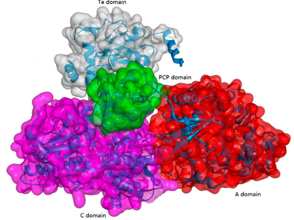
]]>Non-ribosomal peptide synthesis is a universal and critical biochemical process catalyzed by NRPSs in bacteria and fungi, through which a wide array of therapeutically important peptides with highly diverse structures and bioactivities, including penicillin, bleomycin, and cyclosporine, are produced. Biocatalysts capable of NRP synthesis could be divided into two groups (ATP-independent and ATP-dependent enzymes) based on the difference in substrate activation. In the ATP-dependent process, enzymes such as tRNA-dependent ligase can activate the substrate through aminoacyl-adenosine monophosphate. In the ATP-independent process, enzymes such as transacylase use aminoacyl phosphate.

Peptide Synthesis at Creative Peptides
| Services | Description |
| Peptide Synthesis Services | We provide high-quality customized peptide synthesis services, from bulk API peptides, high-throughput library peptides, cosmetic peptides, to array peptides, antigen peptides, and other complex or abnormal peptide sequences. |
| Peptide Modification Services | Creative Peptides offers hundreds of peptide modifications to meet any research need. These modifications can improve the overall stability of peptide, change the structure to better understand biological functions, or enhance the immunogenicity of antibody development and production. |
| Custom Conjugation Service | Creative Peptides provides convenient multiple types of peptide conjugation services to meet the needs of different projects, including activating immune responses, obtaining higher stability and cell penetration. |
ATP-Grasp Enzymes
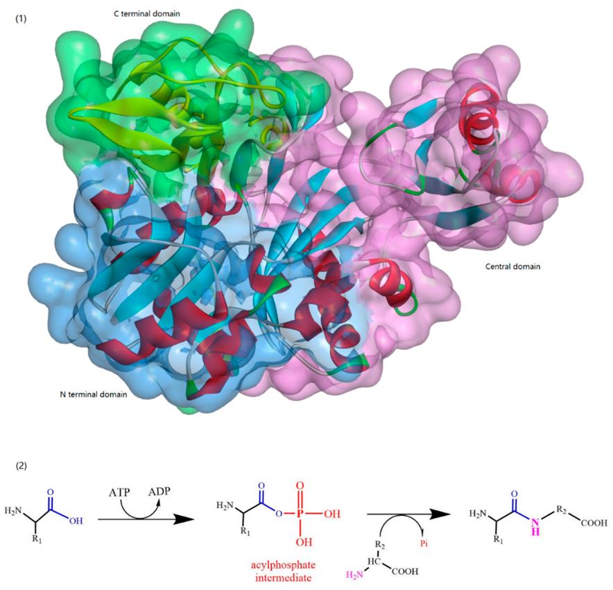
The ATP-grasp enzymes, also known as ATP-dependent carboxylate-amine ligases, activate acids like acylphosphate intermediates and are found in various biological systems such as de novo purine biosynthesis. Notable members of this family include biotin carboxylase, Ddl, and glutathione synthetase. ATP-grasp enzymes usually have a unique structure with three conserved domains, with a nonclassical ATP binding fold enclosing an ATP molecule. Most of these enzymes require an Mg2+ ion coordinated by ATP in the active site.
α-Amino Acid Ester Acyltransferase
Kenzo and colleagues reported an efficient enzymatic method for producing oligopeptides from unprotected amino acids with high yield, using Empedobacter brevis ATCC 14234 strain. An enzyme catalyst (named carboxypeptidase Y) was found in the strain to assist in rapid production of certain oligopeptides. However, details about the amino acid sequence, coding gene sequence or 3D crystal structure were not provided. Isao ABE and team first reported about the cloning and expression of α-amino ester acyl transferases (AETs) from the same and another strain, their amino acid sequences were 35% and 36% identical to the α-amino acid ester hydrolase from Acetobacter pasteurianus. AETs displayed dual action-dipeptidyl peptidase and transferase activities, and was quite specific for both acyl donors and nucleophiles. There have been no studies yet on the 3D structure of AET or its reaction mechanisms.

Enzymes Used for β-Lactam Biosynthesis (β-Lactam Acylases)
β-Lactam antibiotics, which contain a β-lactam ring in their chemical structure and include types such as penicillin, cephalosporins, and thiamycins, are widely used as anti-infective agents and have significant importance in the pharmaceutical industry. The environmentally-friendly and cost-effective synthesis of β-lactam antibiotics via enzymatic means has been increasingly applied in modern pharmaceutical enterprises. β-lactam acylases, which historically process β-lactam antibiotics, can also be used for the creation of semi-synthetic β-lactam antibiotics. Different types of β-lactam acylases such as penicillin acylase (PA), glutaryl acylase (GA), and β-amino acid ester hydrolase (AEH) have been studied in the biosynthesis of β-lactam antibiotics. PAs are generally produced by a variety of microorganisms and are categorized into two types based on their substrate specificity. They are widely used in the industrial production of the active pharmaceutical intermediate 6-aminopenicillanic acid (6-APA) and for the synthesis of semi-synthetic antibiotics, making them potentially useful in the development of novel drugs. Furthermore, PAs can be used in peptide synthesis, resolution of racemic mixtures, and in the production of both achiral and chiral compounds for pharmaceutical intermediates.
Cyanophycinases (CGPases)
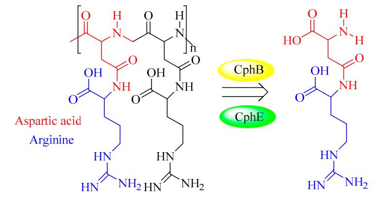
Cyanophycin granule polypeptide (CGP, or multi-L-arginyl-poly) is an intracellular storage polymer found in most cyanobacteria. Equimolar concentrations of arginine and aspartic acid are observed in the aspartic acid backbone, where the arginine moieties are linked to the β-carboxyl group of each aspartic acid through its α-amino group. In most genera of cyanobacteria, the cyanophycin synthetase gene (cphA) has been identified and verified for the synthesis of CGP. In contrast, the intracellular and extracellular degradation of CGP is catalyzed by cyanophycinases (CphB and CphE), which releases dipeptides (β-Asp-Arg). In this respect, β-Asp-Arg can be efficiently synthesized via the simultaneous production of CGP and CGPase, which could be further applied in various fields requiring arginine (Arg) content in feed or food. However, the production and efficient isolation of CGP from various organisms have been successfully established in several recombinant strains, including Escherichia coli, Nicotiana tabacum, Pseudomonas putida, and Pseudomonas alcaligenes DIP1. Therefore, it is very feasible to produce dipeptides (e.g., β-Asp-Arg) via the metabolic engineering of suitable hosts and chemo-enzymatic strategies. For example, successful co-expression of CGP and CGPase in the Nicotiana tabacum plant was recently achieved A further study showed that it is possible to realize the goal of sufficient storage and efficient transport of arginine and β-Asp-Arg dipeptides in this synthetic model.
Reference:
- Wang, T., et al. Strategy for the biosynthesis of short oligopeptides: Green and sustainable chemistry. Biomolecules. 2019, 9(11): 733.
5 X10⁻⁹ M.
In a very detailed study, Cochet-Meilhac & Chambon (1974) demonstrated that the amatoxins bind to RNA polymerase B with a 1:1 stoichiometry, that the affinity of the toxin for the enzyme is very strong with an equilibrium association constant of 3.5 X 10⁻⁸ M-1 at 30 ℃, and that the dissociation is very slow and highly dependent upon temperature and ionic strength. Subsequently, it was shown by Brodner & Wieland (1976), using a carbodiimidecondensation reaction of labeled amatoxin to calf thymus RNA polymerase B, that the 140-kilodalton subunit was a binding site for the amatoxins. Thus, it has been clearly shown that inhibition by a-amanitin is the result of a direct interaction of the toxin with RNA polymerase B.
The process of RNA synthesis catalyzed by DNA-dependent RNA polymerase is quite complex and may be described in several steps as follows: (a) formation of a stable binary complex with the DNA template; (b) RNA chain initiation; (c) translocation and elongation of the nascent RNA; (d) termination and dissociation of the RNA chain. Hence, there are a number of enzyme mechanisms which could possibly be specifically disrupted by a-amanitin. It has been shown that the binding of a-amanitin does not cause a dissociation of the DNA-RNA polymerase B binary complex or the dissociation of the DNA-enzymeRNA ternary complex (Cochet-Meilhac & Chambon, 1974). In the same study, direct evidence was given that the binding of a-amanitin was responsible for the inhibition of RNA chain elongation as catalyzed by RNA polymerase B. The inhibition of chain initiation was suggestedn by indirect experimentation, i.e., by a failure to detect pyrophosphate exchange which is presumably the reverse reaction of phosphodiester bond formation (Krakow & Fronk, 1969). Therefore, it was concluded by Cochet-Meilhac & Chambon (1974) that the binding of amatoxin to RNA polymerase B prevents the formation of phosphodiester bonds.
Reference:
Vaisius, A. C., & Wieland, T. (1982). Formation of a single phosphodiester bond by RNA polymerase B from calf thymus is not inhibited by. alpha.-amanitin. Biochemistry, 21(13), 3097-3101.
]]>
The details of haemopoiesis presented thus far prompt two very important questions. How is the correct balance between stem cell self-renewal and differentiation maintained? And what forces exist that regulate the process of differentiation? The answer to both questions, in particular the latter, is beginning to emerge in the form of a group of cytokines termed ‘haemopoietic growth factors’ (Figure 1). This group includes:
- several (of the previously described) interleukins (ILs) that primarily affect production and differentiation of lymphocytes;
- colony stimulating factors (CSFs), which play a major role in the differentiation of stemderived cells into neutrophils, macrophages, megakaryocytes (from which platelets are derived), eosinophils and basophils;
- erythropoietin, which is essential in the production of red blood cells;
- thrombopoietin, which is essential in the production of platelets.

Figure 1: Major haemopoietic growth factors described to date
GRANULOCYTE-COLONY STIMULATING FACTOR (G-CSF)
G-CSF is also known as pluripoietin and CSF-β. Two slight variants are known, one consisting of 174 amino acids, the other of 177. These are products of alternative splicing. The smaller polypeptide predominates, and also displays significantly greater biological activity than the larger variant. It is synthesized by various cell types, and functions as a growth and differentiation factor for neutrophils and their precursor cells. It also appears to activate mature neutrophils (which are leukocytes capables of ingesting and killing bacteria). G-CSF also appears to act in synergy with additional haemopoietic growth factors to stimulate growth/differentiation of various other haemopoietic progenitor cells. In addition, this cytokine promotes the proliferation and migration of endothelial cells.
MACROPHAGE COLONY-STIMULATING FACTOR (M-CSF)
M-CSF serves as a growth, differentiation and activation factor for macrophages and their precursor cells. It is also known as CSF-1. This cytokine is produced by various cell types. The mature form is a glycoprotein (containing three potential N-linked glycosylation sites), and its 3-D structure is stabilized by multiple disulphide linkages. Three related forms of human M-CSF have been characterized. All are ultimately derived from the same gene and share common C- and N-termini. The largest consists of 522 amino acids with the 406 and 224 amino acid forms lacking different lengths of the internal sequence of the 522 form. The molecular masses of these mature M-CSFs are in the range 45-90 kDa.
GRANULOCYTE-MACROPHAGE COLONY STIMULATING FACTOR (GM-CSF)
GM-CSF is also known as CSF-a or pluripoietin-a. It is a 127 amino acid, single chain, glycosylated polypeptide, exhibiting a molecular mass in the region of 22 kDa. Its 3-D structure exhibits a bundle of four a-helices and a two-stranded antiparallel b-sheet. It thus resembles the other CSFs. GM-CSF is produced by various cells and studies have indicated that its biological activities include:
- Proliferation/differentiation factor of haemopoietic progenitor cells, particularly those yielding neutrophils (a variety of granulocyte) and macrophages, but also eosinophils, erythrocytes and megakaryocytes. In vivo studies also demonstrate this cytokine’s ability to promote haemopoiesis.
- Activation of mature haemopoietic cells, resulting in: enhanced phagocytic activity; enhanced microbiocidal activity; augmented anti-tumour activity; enhanced leukocyte chemotaxis
LEUKAEMIA INHIBITORY FACTOR (LIF)
LIF is an additional haemopoietic growth factor. It is also known as HILDA (human interleukin for DA cells) and hepatocyte stimulatory factor III. It is produced by a range of cell types, including T cells, liver cells and fibroblasts. LIF is a 180 amino acid, heavily glycosylated, 45 kDa protein. It affects both haemopoietic and non haemopoietic tissue, often acting in synergy with other cytokines, particularly IL-3. It stimulates the differentiation of macrophages and promotes enhanced platelet formation. It also prompts synthesis of acute phase proteins by the liver and promotes increased bone resorption. The greatest concentrations of LIF receptors are associated with monocytes, embryonic stem cells, liver cells and the placenta. The receptor complex is composed of two transmembrane glycoproteins: the 190kDa LIFRa-chain which displays affinity for LIF, and a b-chain (gp130), which also forms part of the IL-6 receptor.
ERYTHROPOIETIN (EPO)
EPO is an atypical cytokine in that it acts as a true (endocrine) hormone and is not synthesized by any type of white blood cell. It is encoded by a single copy gene, located on (human) chromosome 7. The gene consists of four introns and five exons. The mature EPO gene product contains 166 amino acids and exhibits a molecular mass in the region of 36 kDa EPO is a glycoprotein, almost 40% of which is carbohydrate. Three N-linked and one O-linked glycosylation site are evident. The O linked carbohydrate moiety apears to play no essential role in the (in vitro or in vivo) biological activity of EPO. Interestingly, removal of the N-linked sugars, while having little effect on EPO’s in vitro activity, destroys its in vivo activity. The sugar components of EPO are likely to contribute to the molecule’s: solubility; cellular processing and secretion; in vivo metabolism. EPO stimulates erythropoiesis by:
- increasing the number of committed cells capable of differentiating into erythrocytes;
- accelerating the rate of differentiation of such precursors;
- increasing the rate of haemoglobin synthesis in developing cells.
THROMBOPOIETIN
Thrombopoietin (TPO) is the haemopoietic growth factor now shown to be the primary physiological regulator of platelet production. Although its existence had been inferred for several decades, its purification from blood proved an almost impossible task, due to its low production levels and the availability of only an extremely cumbersome TPO bioassay. Its existence was finally proved in the mid-1990s when thrombopoietin cDNA was cloned. This molecule is likely to represent an important future therapeutic agent in combating depressed plasma platelet levels, although this remains to be proved by clinical trials. A number of disorders have been identified that are primarily caused by the presence of abnormal platelet levels in the blood. Thrombocythaemia is a disease characterized by abnormal megakaryocyte proliferation, leading to elevated blood platelet levels. In many instances, this results in an elevated risk of spontaneous clot formation within blood vessels. In other instances, the platelets produced are defective, which can increase the risk of spontaneous or prolonged bleeding events.
Reference:
Walsh, G. (2013). Biopharmaceuticals: biochemistry and biotechnology. John Wiley & Sons.
]]>Peptides can regulate the body’s physiological function and provide nutrition to the body. It almost affects the body of all metabolic synthesis. Peptides may be subdivided into oligopeptides and polypeptides. Oligopeptides are made up of two and ten amino acids. And polypeptides are made up of more than ten amino acids. But when up to fifty amino acids, it was called protein. Here, we described the research history of peptide simply.

In 1902, Starling and Bayliss discovered the secretin. They showed that when acid arrived from the stomach it liberated a chemical messenger from the jejunal mucosa, and this messenger traveled through the blood to excite the pancreas to secrete pancreatic juice. This is the first time that human find the peptide. As a result of this discovery, it pioneered the function of peptides in endocrinology. Besides, the Nobel Prize Committee awarded them the Nobel Prize in Physiology.
In 1931, Substance P (SP) was discovered by Ulf von Euler and John H. Gaddum as a tissue extract that caused intestinal contraction in vitro. It is also is a potent vasodilator. Scientists began to pay close attention to the effect of peptides to nervous system and called these substances as neuropeptides.
In 1953, Vincent du Vigneaud studied oxytocin, a hormone that among other things plays a role in sexual intimacy and reproduction among people and mammals. He succeeded in isolating the substance and determining its chemical composition. It became the first peptide hormone to have its sequence of amino acids determined. He also succeeded in producing oxytocin by artificial means.
In 1960, Rita Levi-Montalcini and Stanley Cohen identified a compound, expressed by peripheral cells, that attracted spinal neurons and induced neurite formation. Then they isolated this substance from tumors, snake venom, and mouse salivary glands-all in the face of relentless skepticism from the scientific community. Later, this compound was called nerve growth factor (NGF).
At the end of the 1950s, Robert Bruce Merrifield invent solid-phase peptide synthesis (SPPS).
In 1965, scientists from China completed the synthesis of insulin, which is the world’s first synthetic peptide.
Beginning in 1970, neuropeptide become the research hotspot. Then enkephalin and opioid peptides have been found.
Since the biochemists synthesised peptides by artificial methods for more than 40 years. With the rapid development of molecular biology and biochemical technology, peptide research has made amazing progress.
]]>
When we see the package insert, just like to get the “identity card” of the drugs, so the first we should focus on is the name, including generic name, trade name and chemical name. Actually, this is easy to understand, people also have a formal name , nickname, pseudonym, etc. Then, it’s the picture of drugs—drug structural formula. The formula is extremely important because it can decide all intrinsic properties of the drug. If the structural formula is different then it just like the photo has been changed–“identity” is not the person.
Secondly, drugs also have ID number—[Approval No.], which is unique. It’s available on the website of Food and Drug Administration. We can make fake complaints on those drugs without approval number or the symbol does not match.
Then, generic name is necessary and all the drug manufacturers must use it. That is to say, drugs on the market with a same generic name can come from multiple manufacturers. For example, in terms of Azithromycin tablets, there are 96 manufacturers and 126 manufacturers for injectable Azithromycin tablets, but the trade name is different. During the life, we usually buy a prescription only under the trade name, which might be likely to cause repeated drug use. Thus, it’s necessary to require a careful comparison of the chemical name and formula of drugs. Drugs with the same chemical name and structure decide medicines have the same drug efficiency, pharmacological and toxicological effects.
Why do so many similar drugs have similar generic name? Antibacterial agents could be a good example, such as cephalosporins: cefazolin, cefuroxime, cefotaxime, ceftriaxone, cephalosporins cefepime and lorraine, buserelin acetate, etc. With careful comparison of structure, it’s clear that they have a substantially identical structure, differing only in branches. Therefore, it’s necessary to pay attention to the drugs with common ancestry and family, to avoid side effects and increased drug resistance. In brief, learning the “identity” of drugs can give us a lot of helpful reminders and alerts.
]]>1. It can inhibit the growth hormone, thyroid stimulating hormone, insulin, and pancreatic glucagon secretion.
2. It can inhibit gastric acid secretion stimulated by the test meal and 5 pentagastrin, as well as the release of gastrin and pepsin.
3. It can significantly reduce the splanchnic blood flow, decline portal pressure, blood pressure and collateral circulation, enabling to reduce the blood flow to the liver.
4. It can reduce the secretion of gallbladder and pancreas, as well as internal and external secretion of stomach small intestine, enabling to reduce the activity of pancreatic cell protection.
5. It can inhibit the secretion of glucagon.
6. It can affect the gastrointestinal absorption and nutritional functions to a certain degree, such as liraglutide.

Indications:
It has been used for esophageal varices bleeding due to portal hypertension; gastrointestinal bleeding caused by peptic ulcer, stress ulcer, and erosive gastritis; the prevention and treatment of acute pancreatitis and its complications; pancreas; adjuvant therapy of pancreas, gall bladder, and intestinal fistula; acromegaly, gastrinoma, insulinoma and vasoactive intestinal peptide tumors.
Consumption Usage:
1. Upper gastrointestinal bleeding, mainly indicating the bleeding of esophageal varices: start by intravenous infusion of 250μg (within 3 to 5 minutes), followed by 250μg/hr infusion, and then it should be administered continuously for 48 to 72 hours after bleeding.
2. Pancreas, gall bladder, and intestinal fistula: start with 250μg/h infusion until the fistula closure, and then it’s the successor with 1 to 3 days.
3. Acute pancreatitis: 250μg/h for continuous 72 to 120 hours; prevention of complications of pancreatic surgery for five days in a row; the row ERCP examination should use the product within 2 to 3 hours before the surgery.
Adverse Reactions:
1. Small number of patients may have vertigo, tinnitus, flushing;
2. Injection of this product faster than 50μg/min will produce nausea and vomiting.
Precautions:
1. Banned from the allergy, as well as pregnant and lactating women.
2. Administration at the beginning might cause a temporary decline in blood sugar, so insulin-dependent diabetics should check the blood sugar every 3 to 4 hours.
3. It can be used to extend the hypnotic effecting time of hexobarbital and the effect might aggravate the role of pentene tetrazole. Thus, the two products cannot be used simultaneously.
4. The product should be administered alone, indicating this product should not be administered in combination with other drugs combined.
5. Arterial bleeding is not a somatostatin indication.
Specification:
Injection: 250μg, 3mg
Modern scientific advances in various diseases have been benefited from high-level peptide products, such as deslorelin acetate, GLP-1 (7-36) amide acetate, teduglutide, and so on. That’s why increasing researchers are focusing on peptide studies!
]]>