Basic Principles of SPPS
Peptide synthesis involves a large number of repetitive steps, and the use of a solid support offers significant advantages. In such a system, high concentrations of excess reagents can drive coupling reactions to completion, while surplus reagents and by-products can be separated from the insoluble growing peptide chain (resin) simply by filtration and washing. All operations are carried out in the same solvent, eliminating the need for material transfer.
- Anchoring of the initial amino acid: The initial amino acid is anchored to the resin through its C-terminus, forming either an ester bond or an amide bond.
- Linear assembly: The peptide chain is elongated stepwise from the C-terminus to the N-terminus through condensation reactions.
- Protecting-group strategy: Permanent protecting groups are used for side chains to improve stability; non-side-chain amino acid protection is generally achieved using carbamate-type protecting groups, which can be removed under mild conditions, thereby avoiding racemization caused by overly harsh conditions.
- Cyclic operations: Coupling, washing to remove excess reagents, deprotection, and initiation of the next coupling cycle.
- Final cleavage: The peptide chain is cleaved from the resin while simultaneously removing the side-chain protecting groups.
Fmoc/t-Bu Strategy in Solid-Phase Synthesis
Two major protecting-group strategies are mainly used in solid-phase synthesis: Boc/Bzl and Fmoc/t-Bu. The former requires deprotection under relatively strong acidic conditions, such as TFA, and is mainly used for side-chain protection. Strong acidic conditions often damage sensitive amino acid sequences; therefore, the Fmoc/t-Bu strategy is generally adopted for amino acid protection, in which Fmoc is base-labile and t-Bu is weak-acid-labile. Solid-phase synthesis methods using the Fmoc-based strategy have become the preferred approach for routine peptide synthesis.
Solid Supports
Solid supports can be characterized by both the “matrix polymer” and the “linker molecule,” although the term “resin” is often incorrectly used to refer to the entire system. In solid-phase chemistry, the matrix polymer is as important as the liquid-phase environment. At present, there are well over one hundred commercially available resins, some of which are equipped with the same linker molecules. Therefore, the most suitable linker must be selected carefully during synthesis.
Cross-linked polystyrene (PS)-based resins are commonly used solid supports for SPPS. Resin beads with a particle size distribution of 200-400 mesh (approximately 50 μm in diameter) and a loading of 0.5-0.8 mmol/g exhibit good swelling properties in DMF and DCM, which promotes diffusion of reactants into the polymer matrix and ensures adequate access to linker sites buried within the beads. For longer peptide chains (more than 25 amino acids) or sequences that are more difficult to synthesize, lower-loading resins of 0.1-0.2 mmol/g should be used. Cross-linked polyamide (PA)-based resins and polystyrene-polyethylene glycol (PS-PEG) composite resins are more hydrophilic solid supports. Their microscopic and macroscopic physical properties differ from those of PS resins. These supports generally have lower loading capacities and may serve as alternatives to PS for the synthesis of complex sequences and large peptides.
Resin Handling
Why must resins be treated before use? This is analogous to textile dyeing and finishing. Because cotton fibers have irregular oval cross-sections, they must be swollen before dyeing to increase the surface area so that dyes can penetrate effectively. Resin is a polymeric material with long chains that may become entangled or structurally collapsed, and its reactive sites are located inside the resin. Therefore, swelling treatment before use is essential, and the reaction conditions must also be taken into consideration.
Selection of the Reactor
SPPS reaction vessel: The reactor is generally made of glass and equipped with a PTFE or glass frit syringe. The size of the reactor should be selected according to the amount of resin used.
| Container Length (cm) | Container Diameter (cm) | Maximum Resin Weight (g) | Working Volume (mL) |
| 5 | 2 | 0.5 | 10 |
| 11 | 2.6 | 2 | 40 |
| 15 | 3.4 | 4 | 90 |
SPPS Reactor Types
Selection of Solvents
Ninety-nine percent of the coupling sites are not located on the surface of the resin beads but rather inside the resin. Therefore, resin beads bearing the growing peptide chain must be fully swollen to ensure optimal penetration of the protected activated amino acids into the polymer matrix, thereby improving coupling efficiency.
The basic swelling procedure is as follows: before starting solid-phase synthesis, place the resin in a suitable solvent such as DMF or DCM and allow it to swell for 20-30 minutes. For cross-linked polystyrene (PS)-based resin beads, DCM provides the best swelling characteristics. During the coupling step, however, DMF or NMP is preferred because these solvents provide better solubility for the reactants. Alcohols and water are not suitable for cross-linked polystyrene (PS)-based resins. However, in subsequent washing steps, methanol or isopropanol may be used to shrink the resin beads and expel entrapped excess reactants. After this treatment, the beads must be reswollen with DCM and DMF. Resin swelling procedure: Place the resin in a suitable reactor, add DCM until the resin beads are fully immersed, and gently mix the resin suspension with a PTFE stirring rod. Allow it to swell for 20-30 minutes, then remove the solvent by vacuum filtration.
Stirring and Mixing
During SPPS, reaction kinetics are governed primarily by diffusion phenomena; therefore, vigorous stirring of the reaction vessel is unnecessary. In addition, most resin beads used in peptide synthesis are relatively fragile, and strong agitation can damage or fracture them. Gentle mixing may be achieved using the rotor of a rotary evaporator or by employing devices capable of mild motion such as rocking or vortex-like mixing.
Washing
Washing is used to remove soluble by-products and excess reagents generated during coupling and deprotection. The basic washing procedure involves introducing solvent into the reactor and then removing it under vacuum; when necessary, the resin may be stirred gently in the washing solvent with a stirring rod.
Standard washing and reswelling procedure: Add DMF to the reaction vessel, allow it to stand for 10 s, then remove the solvent by vacuum filtration, carefully rinsing all parts of the reactor with DMF. Remove the solvent under vacuum, then add fresh solvent again for washing, repeating the operation 1-2 times. After washing is complete, wash the resin with methanol using a procedure similar to the DMF washing step, repeating 1-2 times. Then switch to DCM for washing 1-2 times, followed by DMF washing 1-2 times.
Linkers and Resin Selection in Fmoc Solid-Phase Synthesis
The first step in SPPS is to anchor the C-terminal amino acid residue (with its N-terminus protected) to the solid support through either an ester bond or an amide bond, depending on whether the target peptide has a C-terminal carboxylic acid or a C-terminal amide group. At present, most linker molecules are commercially available and preloaded onto different matrices, including polystyrene (PS), polyamide (PA), and polystyrene-polyethylene glycol (PS-PEG) composite matrices. C-terminal peptide acids are generated through ester linkages, whereas C-terminal peptide amides are generated through amide linkages. These preloaded linkers simplify experimental preparation and ensure batch-to-batch consistency. Different matrices have different characteristics: PS matrices are highly hydrophobic and suitable for routine peptide synthesis; PS-PEG matrices are more hydrophilic and advantageous for long-peptide synthesis; and polyamide (PA) is used for specialized applications.
Peptide Amides
For the synthesis of C-terminal peptide amides, the following resins are commonly used. The attachment of the first amino acid to these resins is basically consistent with the Fmoc synthesis strategy, and under TFA treatment these resins leave an amide bond upon cleavage to complete the release of the peptide (the resin functions similarly to a protecting group). Because these resins contain free amino groups, they are usually Fmoc-protected and must be deprotected before use. It should be noted that when amino acids with substantial steric hindrance at the C-terminus are coupled to the resin, incomplete conversion may occur, and a second coupling step may be required.
Peptide Acids
Compared with the nucleophilicity of amino groups, hydroxyl groups are less nucleophilic. Therefore, anchoring amino acids onto a solid support in the form of esters is more challenging. It is recommended to directly purchase resins preloaded with the protected C-terminal amino acid to avoid issues such as epimerization, dipeptide formation, and substitution-level variability. When synthesizing C-terminal peptide acids using the Fmoc/tBu strategy, the anchoring reaction must be carried out under anhydrous conditions, and amino acids containing moisture must be dried in advance.
Hydroxyl Resins
Among hydroxyl resins, the less sterically hindered Wang resin more readily forms ester bonds, and the symmetrical anhydride method is commonly used. If anchoring is difficult, the esterification step may be repeated with fresh reagents; some amino acid derivatives may require three esterification cycles to achieve satisfactory results. After anchoring is completed, the unreacted hydroxyl groups on the resin are recommended to be capped with benzoic anhydride or acetic anhydride. Standard operating procedure for hydroxyl resins: First place the resin in the reactor and swell it with a suitable solvent. Meanwhile, dissolve the target Fmoc-protected amino acid (10.0 eq.) in DCM at 0°C, add DIC (5.0 eq.) and allow activation for 10 minutes; DMF may be added if necessary to aid dissolution. Transfer the activated solution to the resin, add DMAP/DMF to catalyze the reaction, and stir for 1 hour; then wash once each with DMF and DCM. Dry under vacuum for 18 hours and determine the loading by the Fmoc release assay. If the loading is below 70%, the esterification step should be repeated.
Once the target loading is achieved, cap the residual hydroxyl groups with benzoic anhydride/acetic anhydride (5 eq.) and pyridine (1 eq.) in DMF for 30 minutes. After final washing and Fmoc deprotection, peptide-chain elongation may begin. This procedure ensures coupling efficiency through low-temperature activation, strict stoichiometric control, and loading verification. Key steps include activation monitoring, catalytic enhancement, and hydroxyl capping to prevent by-product formation.
Trityl-Series Resins
These resins exhibit high acid lability, and the steric hindrance of their linker groups can significantly suppress diketopiperazine (DKP) formation. They are particularly recommended for the synthesis of peptides with C-terminal proline (Pro) or glycine (Gly). These resins can be cleaved under extremely mild acidic conditions, thereby protecting both the peptide segment and the resin. Commercial products are generally supplied as chlorotrityl resin or trityl alcohol precursors.
Typical procedure for trityl resin: Place 1 g of trityl resin (loading 1.0-2.0 mmol chloride/g) into an SPPS reactor and allow it to swell. Add 3 equivalents of the Fmoc amino acid and 7.5 equivalents of DIPEA, then stir at room temperature in anhydrous DCM for 30-60 minutes. Wash the resin sequentially with DMF 2-3 times, then add 10 mL of a DCM/MeOH/DIPEA mixture to cap the residual chloride groups, and continue the reaction for 15 minutes. Finally, wash three times each with DMF and DCM, dry under vacuum, and determine the loading by the Fmoc release assay. Through this procedure, precise control of the amino acid-to-DIPEA ratio, together with methanol quenching, ensures single-site attachment. It should be noted that the activity of trityl chloride must be maintained under anhydrous conditions.
]]>Structural Features and Pharmacological Advantages
Macrocyclic peptides are a class of peptide molecules characterized by a ring-like structure, typically composed of 5-17 amino acid residues with a molecular weight ranging from 500 to 2000 Da. Positioned between small-molecule compounds and large biological therapeutics, they combine the structural and functional advantages of both, making them a promising direction in peptide drug discovery. The cyclic architecture endows these peptides with exceptional target-binding affinity and biological stability, enabling them to effectively modulate protein-protein interactions (PPIs) and intrinsically disordered proteins (IDPs)—targets traditionally considered “undruggable.” These features highlight their vast potential in the development of orally available peptide drugs.
Enhanced Bioactivity, Stability, and Selectivity
Compared with linear peptides, macrocyclic peptides demonstrate higher target specificity and biological activity, allowing for potent therapeutic effects at lower doses while minimizing off-target interactions and systemic toxicity. Their rigid cyclic structure improves resistance to gastric acid and proteolytic enzymes, prolonging in vivo half-life and enhancing pharmacodynamic persistence. Moreover, macrocyclic peptides exhibit remarkable structural diversity and design flexibility, allowing chemical modifications to meet diverse therapeutic needs. With low immunogenicity, strong target selectivity, and minimal impact on normal cells, they are emerging as safe and effective candidates for next-generation precision therapeutics. Continuous advancements in delivery technologies further enhance their clinical feasibility.
With their superior pharmacological profiles and evolving synthetic technologies, macrocyclic peptides have become one of the hottest frontiers in peptide drug research. They hold the promise of achieving precise target engagement, improved safety profiles, and optimized efficacy, positioning themselves as a cornerstone in the future of innovative therapeutic strategies.
Clinical Pipeline of Macrocyclic Peptide Drugs
The number of macrocyclic peptide drugs entering clinical development has been steadily increasing (Table 1), with orally administered macrocyclic peptides emerging as a major focus area. Since the approval of Cyclosporine A by the FDA in 1983, macrocyclic peptide therapeutics have made remarkable advances across multiple therapeutic fields. However, orally bioavailable macrocyclic peptides remain relatively rare. With the rapid emergence of new technologies, the development of oral peptide therapeutics is expected to achieve significant breakthroughs in the coming years. Several oral macrocyclic peptide candidates have now advanced into clinical trials, with representative examples including the following:
01. MK-0616 (Merck & Co.)
MK-0616 is an oral PCSK9 inhibitor currently in Phase III clinical trials. Designed as a macrocyclic peptide capable of binding to PCSK9 (Proprotein Convertase Subtilisin/Kexin Type 9), MK-0616 aims to reduce low-density lipoprotein cholesterol (LDL-C) levels. In contrast to current PCSK9-targeting antibody therapies that require subcutaneous injection, MK-0616’s oral formulation is expected to greatly improve patient adherence and reduce treatment costs. Although the peptide itself has limited permeability, the inclusion of an absorption enhancer allows an oral bioavailability of approximately 2%. On June 9, 2025, Merck announced that both pivotal Phase III trials—CORALreef HeFH and CORALreef AddOn—achieved their primary endpoints in patients with hypercholesterolemia, marking a major milestone for oral peptide drug development.
02. JNJ-2113 (Johnson & Johnson)
JNJ-2113 is an oral IL-23 receptor antagonist currently in Phase III clinical development. The program originated from a collaboration between Protagonist Therapeutics and Johnson & Johnson, established in 2017, with J&J leading subsequent development and commercialization. In November 2024, JNJ-2113 achieved positive topline results in its pivotal ICONIC-LEAD Phase III trial, demonstrating robust efficacy and oral potential in patients with moderate-to-severe plaque psoriasis (PsO), including adolescents aged 12 years and older. If successfully approved, JNJ-2113 is poised to become the first oral peptide drug targeting the IL-23 receptor, representing a transformative step in the treatment of chronic inflammatory diseases.
03. LUNA18 (Chugai Pharmaceutical)
LUNA18 is a KRAS inhibitor currently in Phase I clinical trials in Japan and the United States. In preclinical studies, LUNA18 demonstrated oral bioavailability ranging from 21% to 47% across multiple species (mice, rats, monkeys, and dogs), without the need for permeability enhancers—indicating excellent membrane penetration and drug-like properties. Moreover, dose-dependent antitumor activity was observed in animal models, underscoring its strong therapeutic potential as a next-generation macrocyclic peptide-based KRAS inhibitor.
| Drug Name | Highest Clinical Phase | Source Type | Clinical Trial ID | Route of Administration |
| AP301 | Phase III | Natural product derivative | NCT04551300 | Oral |
| BMS-986229 | Phase III | mRNA display | NCT04161781 | Injectable |
| JNJ-2113 | Phase III | Phage display | NCT05364554 | Oral |
| MK-0616 | Phase III | mRNA display | NCT05952896 | Oral |
| ORMD-0801 | Phase III | Natural product derivative | NCT04817215 | Oral |
| PL9643 | Phase III | Natural product derivative | NCT05201170 | Ophthalmic |
| Plitidepsin | Phase III | Natural product source | NCT01102426 | Injectable |
| Balixafortide | Phase III | Natural product derivative | NCT03786094 | Injectable |
| Rusfertide | Phase III | Natural product derivative | NCT04202965 | Injectable |
| BT8009 | Phase II/III | Phage display | NCT04561362 | Injectable |
| VT1021 | Phase II/III | Natural product derivative | NCT03970447 | Injectable |
| ALRN-6924 | Phase II | Stapled peptide design | NCT02264613 | Injectable |
| AMY-101 | Phase II | Phage display | NCT03694444 | Injectable |
| AZP-3813 | Phase II | mRNA display | Injectable | |
| Certepediol | Phase II | Phage display | NCT03517116 | Injectable |
| Dolcamatide | Phase II | Natural product derivative | NCT01983306 | Oral |
| PL8177 | Phase II | Natural product derivative | NCT05466890 | Oral |
| THR-149 | Phase II | Phage display | NCT04527107 | Injectable |
| TE-232 | Phase II | Natural product derivative | NCT00422786 | Injectable |
| BHV-1100 | Phase I/II | mRNA display | NCT04634435 | Injectable |
| BT1718 | Phase I/II | Phage display | NCT03486730 | Injectable |
| FOG-001 | Phase I/II | Phage display | NCT05919264 | Injectable |
| Lonodelestat | Phase I/II | NCT03748199 | Inhalation | |
| BT5528 | Phase I/II | Phage display | NCT04180371 | Injectable |
| BT7480 | Phase I/II | Phage display | NCT05163041 | Injectable |
| LUNA18 | Phase I | mRNA display | NCT05012618 | Oral |
Cyclic Peptides at Creative Peptides
| CAT# | Product Name | M.W | Molecular Formula |
| 10-101-103 | Vancomycin | 1449.25 | C66H75Cl2N9O24 |
| 10-101-104 | Teicoplanin | 1879.66 | C88H95Cl2N9O33 |
| 10-101-112 | Bremelanotide | 1025.18 | C50H68N14O10 |
| 10-101-169 | Pasireotide | 1047.20624 | C58H66N10O9 |
| 10-101-186 | Romidepsin | 540.69584 | C24H36N4O6S2 |
| 10-101-325 | Semaglutide | 4113.57 | C187H291N45O59 |
| 10-101-62 | Ziconotide | 2639.13 | C102H172N36O32S7 |
| 10-101-78 | Dalbavancin | 1816.69 | C88H100Cl2N10O28 |
| AF083 | Polymyxin B | ||
| MFP-041 | Rezafungin | 1226.4 | C63H85N8O17 |
| R04030 | Cyclo(-Arg-Gly-Asp-D-Phe-Val) | 574.64 | |
| R1574 | Octreotide | 1019.24 | C₄₉H₆₆N₁₀O₁₀S₂ |
| R1812 | Lanreotide | 1096.33 | C54H69N11O10S2 |
| R1824 | Cyclo(RGDyK) | C31H43F6N9O12 | |
| R2018 | Capreomycin | ||
| R2029 | Enviomycin | 685.69 | C26H43N11O11 |
| R2052 | Zilucoplan | (C2H4O)nC126H186N24O32 | |
| R2238 | Telavancin | 1755.6 | C80H106Cl2N11O27P |
| R2239 | Oritavancin | 1793.1 | C86H97Cl3N10O26 |
| R2240 | Bacitracin | 1422.69 | C66H103N17O16S |
Cell-penetrating peptides (CPPs) are short peptides capable of entering cells without disrupting membrane integrity. They typically consist of 5 to 30 amino acids and are rich in basic amino acids, including arginine and lysine. Under physiological conditions, CPPs share two common characteristics: a positive charge and amphiphilicity. In recent years, CPPs have been widely applied in drug delivery systems.
The recognized mechanisms by which CPPs penetrate cell membranes fall into two categories: endocytosis and direct translocation. Endocytosis is an energy-dependent process and includes three main pathways: macropinocytosis, caveolae-mediated endocytosis (CvME), and clathrin-mediated endocytosis (CME). In contrast, direct translocation is an energy-independent process facilitated by electrostatic interactions between the positively charged CPPs and the negatively charged membrane components and phospholipid bilayer, promoting direct membrane permeation. Several models have been proposed for direct penetration, including the barrel-stave model, carpet model, toroidal pore model, and inverted micelle model.
A summary of the cell-penetrating peptide used in drug delivery is as follows:
1. Polyarginine
Single-letter code: H2N-RRRRRRRRR-OH
Three-letter code: H2N-Arg-Arg-Arg-Arg-Arg-Arg-Arg-Arg-Arg-OH
Number of amino acids: 9
Molecular formula: C54H110N36O10
Average molecular weight (MW): 1423.69
2. Transportan 10 (TP10)
Single-letter code: H2N-AGYLLGKINLKALAALAKKIL-OH
Three-letter code: H2N-Ala-Gly-Tyr-Leu-Leu-Gly-Lys-Ile-Asn-Leu-Lys-Ala-Leu-Ala-Ala-Leu-Ala-Lys-Lys-Ile-Leu-OH
Number of amino acids: 21
Molecular formula: C104H184N26O24
Average molecular weight (MW): 2182.73
3. Tumor-Lineage Homing CPP
Single-letter code: H2N-RLYMRYYSPTTRRYG-OH
Three-letter code: H2N-Arg-Leu-Tyr-Met-Arg-Tyr-Tyr-Ser-Pro-Thr-Thr-Arg-Arg-Tyr-Gly-OH
Number of amino acids: 15
Molecular formula: C89H135N27O23S1
Average molecular weight (MW): 1983.26
4. dNP2
Single-letter code: H2N-CKIKKVKKKGRKKIKKVKKKGRK-OH
Three-letter code: H2N-Cys-Lys-Ile-Lys-Lys-Val-Lys-Lys-Lys-Gly-Arg-Lys-Lys-Ile-Lys-Lys-Val-Lys-Lys-Lys-Gly-Arg-Lys-OH
Number of amino acids: 23
Molecular formula: C125H245N43O24S1
Average molecular weight (MW): 2766.62
5. RVRR
Single-letter code: H2N-RVRR-OH
Three-letter code: H2N-Arg-Val-Arg-Arg-OH
Number of amino acids: 4
Molecular formula: C23H47N13O5
Average molecular weight (MW): 585.70
6. L-penetratin
Single-letter code: H2N-RQIKIWFQNRRMKWKK-OH
Three-letter code: H2N-Arg-Gln-Ile-Lys-Ile-Trp-Phe-Gln-Asn-Arg-Arg-Met-Lys-Trp-Lys-Lys-OH
Number of amino acids: 16
Molecular formula: C104H168N34O20S1
Average molecular weight (MW): 2246.73
7. Activable CPP (ACPP)
Single-letter code: H2N-CRRRRRRRRGGGPKKKKKK-OH
Three-letter code: H2N-Cys-Arg-Arg-Arg-Arg-Arg-Arg-Arg-Arg-Gly-Gly-Gly-Pro-Lys-Lys-Lys-Lys-Lys-Lys-OH
Number of amino acids: 19
Molecular formula: C98H191N49O20S1
Average molecular weight (MW): 2407.95
8. LH2
Single-letter code: Ac-LHHLCHLLHHLCHLAG-CONH2
Three-letter code: Ac-Leu-His-His-Leu-Cys-His-Leu-Leu-His-His-Leu-Cys-His-Leu-Ala-Gly-CONH2
Number of amino acids: 16
Molecular formula: C85H131O17N29S2
Average molecular weight (MW): 1895.26
9. Cancer Cell Line Specific CPP
Single-letter code: H2N-RLYMRYYSPTTRRYG-OH
Three-letter code: H2N-Arg-Leu-Tyr-Met-Arg-Tyr-Tyr-Ser-Pro-Thr-Thr-Arg-Arg-Tyr-Gly-OH
Number of amino acids: 15
Molecular formula: C89H135N27O23S1
Average molecular weight (MW): 1983.26
10. T2
Single-letter code: H2N-FKKFFRKLL-OH
Three-letter code: H2N-Phe-Lys-Lys-Phe-Phe-Arg-Lys-Leu-Leu-OH
Number of amino acids: 9
Molecular formula: C63H99N15O10
Average molecular weight (MW): 1226.55
Cell-penetrating Peptides (CPPs) at Creative Peptides
- Antennapedia Peptides
- Chimeric Peptides
- HIV-TAT Family
- Nuclear Localization Peptides
- Oligoarginine Peptides
People also like
- Cell Targeting Peptides: Definition, Classification and Application
- Cell-penetrating peptides (CPPs) for Diagnostics and Imaging
- Cell-penetrating Peptides (CPPs) for Gene Therapy
- Cell-penetrating Peptides (CPPs) for Drug Delivery
- Cyclic Cell Penetrating Peptides: Definition, Structure, Mechanism and Applications
- The Design of Cell Penetrating Peptides
- Classes and Applications of Cell-Penetrating Peptides
1. RGD
The tripeptide RGD can specifically bind to integrin receptors. Integrins are composed of two subunits (α and β). The integrin family, especially αVβ3, plays a crucial role in tumor angiogenesis and metastasis. During angiogenesis, these receptors are overexpressed on endothelial cells but are barely detectable in most normal organs. Therefore, they are widely used in diagnostic imaging.
Single-letter code: H2N-RGD-OH
Three-letter code: H2N-Arg-Gly-Asp-OH
Number of amino acids: 3
Molecular formula: C12H22N6O6
Average molecular weight (MW): 346.34
2. Bombesin (BBN) / Gastrin-Releasing Peptide (GRP)
BBN and its related peptides from amphibians form a neuropeptide family with multiple physiological effects, such as exocrine and endocrine secretion, thermoregulation, sucrose regulation, and cell growth. Bombesin-like peptide receptors have four subtypes: neuromedin B receptor, bombesin receptor subtype 3, gastrin-releasing peptide receptor (GRPR), and bombesin receptor subtype 4. These receptors are overexpressed in various tumors, including breast cancer, ovarian cancer, and gastrointestinal stromal tumors.
| CAT# | Product Name |
| B07004 | Bombesin (8-14) |
| B07010 | [Lys3]-Bombesin |
| B07011 | [Tyr4, D-Phe12]-Bombesin |
| B07015 | (D-Cys6,Asn7,D-Ala11,Cys14)-Bombesin (6-14) |
| B07016 | (D-Phe12)-Bombesin |
| B07017 | (D-Phe12,Leu14)-Bombesin |
| B07019 | (D-Phe6,Leu-NHEt13,des-Met14)-Bombesin (6-14) |
| B07020 | (D-Tyr6,betaPhe11,Phe13,Nle14)-Bombesin (6-14) (free acid) |
| B07022 | (Tyr4)-Bombesin |
| B07023 | Cyclo(-D-Phe-His-Trp-Ala-Val-Gly-His-Leu-Leu) |
| B07024 | Biotin – Bombesin |
| B07025 | Biotin – LC – LC – Bombesin |
| B07026 | Bombesin, FAM – labeled |
Gastrin-Releasing Peptide (GRP)
3. Somatostatin (SST) Peptides
Somatostatins (SSTs) are naturally occurring cyclic peptide hormones composed of either 14 or 28 amino acids. They inhibit the secretion of insulin, glucagon, and several other hormones. Their biological effects are mediated by binding to specific high-affinity somatostatin receptors (SSTRs), of which there are five subtypes: SSTR1 to SSTR5. These receptors are overexpressed in a variety of neuroendocrine tumors, such as pancreatic neuroendocrine tumors, small cell lung cancer, and thyroid carcinoma. Synthetic somatostatin analogs are widely used in imaging and therapy of tumors that express these receptors.
| CAT# | Product Name |
| 10-101-169 | Pasireotide |
| 10-101-26 | Octreotide Acetate |
| 10-101-44 | Vapreotide Acetate |
| M34140635H | [Nal3]Octreotide acetate |
| M34140636H | TETA-Octreotide acetate |
| M34140637H | NOTA-Octreotide trifluoroacetate |
| M34140643H | DOTA-Lanreotide acetate |
| M34140653H | [Tyr3,Lys5(Boc)]octreotide acetate |
| M34140655H | [Lys5(Boc)]lanreotide acetate |
| O1003 | ([ring-D5]Phe3)-Octreotide |
| S07018 | (D-2-Nal5,Cys6·11,Tyr7,D-Trp8,Val10,2-Nal12)-Somatostatin-14 (5-12) amide |
| S07041 | Tyr-(D-Dab4,Arg5,D-Trp8)-cyclo-Somatostatin-14 (4-11) |
| S07043 | Vapreotide |
4. Vasoactive Intestinal Peptide (VIP)
VIP is a neuropeptide consisting of 28 amino acids. It not only promotes vasodilation but also stimulates cell growth and proliferation through cell surface receptor-mediated signaling pathways. Its effects are primarily regulated by two receptor subtypes, VPAC1 and VPAC2. VIP receptors are highly expressed in a variety of tumors, including but not limited to brain tumors, pancreatic adenocarcinomas, and neuroendocrine tumors. Notably, VIP is also considered a potential therapeutic agent; however, toxicity may occur even at sub-microgram doses.
Single-letter sequence: H2N-HSDAVFTDNYTRLRKQMAVKKYLNSILN-OH
Three-letter sequence: H2N-His-Ser-Asp-Ala-Val-Phe-Thr-Asp-Asn-Tyr-Thr-Arg-Leu-Arg-Lys-Gln-Met-Ala-Val-Lys-Lys-Tyr-Leu-Asn-Ser-Ile-Leu-Asn-OH
Number of amino acids: 28
Molecular formula: C147H237N43O43S1
Average molecular weight (MW): 3326.78
Vasoactive Intestinal Peptides (VIPs)
- Cholecystokinin (CCK) / Gastrin Peptide
Cholecystokinin (CCK) and gastrin are structurally and functionally related peptides that play multiple physiological roles in the gastrointestinal tract and central nervous system. They share an identical sequence in their bioactive region, differing only in the site of tyrosine sulfation—position 6 in gastrin and position 7 in CCK. To date, three CCK receptors have been identified: CCK1, CCK2, and CCK2i4sv, all of which belong to the G protein-coupled receptor (GPCR) superfamily. Among them, the CCK2/gastrin receptor is frequently implicated in human cancers, such as ovarian stromal tumors and astrocytomas.
Single-letter sequence: H2N-D-sTyr-MGWMDF-OH
Three-letter sequence: H2N-Asp-Tyr(SO3H)-Met-Gly-Trp-Met-Asp-Phe-OH
Number of amino acids: 8
Molecular formula: C49H61N9O17S3
Average molecular weight (MW): 1144.25
6. Alpha-Melanocyte-Stimulating Hormone (α-MSH)
Alpha-Melanocyte-Stimulating Hormone (α-MSH) is a linear tridecapeptide primarily responsible for regulating skin pigmentation. α-MSH and its analogs exhibit extremely high binding affinity to the melanocortin-1 receptor (MC-1R), which is expressed in over 80% of human melanoma metastases. Therefore, α-MSH is widely used as a carrier for targeted imaging and radiotherapy of melanoma.
Single-letter sequence: H2N-SYSMEHFRWGKPV-OH
Three-letter sequence: H2N-Ser-Tyr-Ser-Met-Glu-His-Phe-Arg-Trp-Gly-Lys-Pro-Val-OH
Number of amino acids: 13
Molecular formula: C75H106N20O19S1
Average molecular weight (MW): 1623.83
Melanocyte-Stimulating Hormone (MSH)
| CAT# | Product Name |
| M09001 | a-MSH, amide |
| M09002 | a-MSH, Free Acid |
| M09003 | [Des-Ac] a-MSH, amide |
| M09004 | g-MSH |
| M09005 | g-1-MSH, amide |
| M09006 | MSH Release Inhibiting Factor, amide |
| M09007 | b-MSH, porcine |
| M09009 | [Lys0]-γ-1-MSH (41-58), amide |
| M09010 | Melanocyte Associated Antigen gp 100 (17-25) |
| M09011 | Melanotropin-Potentiating Factor, MPF |
| M09012 | RAB38/NY-MEL-1 (50-58) |
| M09013 | Acetyl-(D-Val13)-a-MSH (11-13) |
7. Neurotensin (NT)
Neurotensin (NT) is a peptide consisting of 13 amino acids that acts on neurotensin receptors, which have been identified in various tumors, such as ductal pancreatic adenocarcinoma, small-cell lung carcinoma, and medullary thyroid carcinoma. Thus, NT is considered a highly promising candidate for cancer imaging applications.
Single-letter sequence: Pyr-LYENKPRRPYIL-OH\
Three-letter sequence: Pyr-Leu-Tyr-Glu-Asn-Lys-Pro-Arg-Arg-Pro-Tyr-Ile-Leu-OH
Number of amino acids: 12
Molecular formula: C78H121N21O20
Average molecular weight (MW): 1672.92
Neurotensins and Related Peptides
| CAT# | Product Name |
| N05044 | (Dab9)-Neurotensin (8-13) |
| N05045 | (D-Trp11)-Neurotensin |
| N05046 | (D-Tyr11)-Neurotensin |
| N05048 | (Lys9,Trp11,Glu12)-Neurotensin (8-13) (Cyclic Analog) |
| N05049 | Acetyl-Neurotensin (8-13) |
| N05050 | Boc-(Lys9)-Neurotensin (9-13)-methyl ester |
| N05051 | Neurotensin (1-11) |
| N05052 | Neurotensin (1-6) |
| N06001 | (Lys8,Lys9)-Neurotensin (8-13) |
| N06002 | [Lys8,Asn9] Neurotensin LANT-6 (8-13) |
| N06003 | [Trp11] Neurotensin (8-13) |
| N06004 | [Trp11]-Neurotensin |
| N06006 | Neurotensin (1-8) |
| N06008 | Neurotensin (9-13) |
| N06009 | [Gln4]-Neurotensin |
8. T140
T140 is a peptide composed of 14 amino acids and one disulfide bond. It is an antagonist of chemokine receptor 4 (CXCR4). Its derivatives have been widely used as imaging agents targeting CXCR4.
Single-letter sequence: H2N-RR-2Nal-CYRKkPYR-Cit-CR-OH
Three-letter sequence: H2N-Arg-Arg-2Nal-Cys-Tyr-Arg-Lys-DLys-Pro-Tyr-Arg-Cit-Cys-Arg-OH
Number of amino acids: 14
Molecular formula: C90H143N33O18S2
Average molecular weight (MW): 2039.44
9. Exendin-4
Exendin-4 is a peptide hormone consisting of 39 amino acids. It shares 50% sequence homology with the natural glucagon-like peptide-1 (GLP-1) and has been identified as an agonist of the GLP-1 receptor. Exendin-4 has been clinically used for the treatment of insulinomas and type 2 diabetes.
Single-letter sequence: H2N-HGEGTFTSDLSKQMEEEAVRLFIEWLKNGGPSSGAPPPS-OH
Three-letter sequence: H2N-His-Gly-Glu-Gly-Thr-Phe-Thr-Ser-Asp-Leu-Ser-Lys-Gln-Met-Glu-Glu-Glu-Ala-Val-Arg-Leu-Phe-Ile-Glu-Trp-Leu-Lys-Asn-Gly-Gly-Pro-Ser-Ser-Gly-Ala-Pro-Pro-Pro-Ser-OH
Number of amino acids: 39
Molecular formula: C184H281N49O61S1
Average molecular weight (MW): 4187.56
| CAT# | Product Name |
| 10-101-83 | Exendin (9-39) Acetate |
| E10001 | Exendin (9-39) |
| E10005 | Exendin-4 (3-39) |
| E10007 | Exendin (10-39) |
| E10008 | Exendin (4-39) |
| E10009 | Exendin (5-39) |
| E10010 | Exendin (7-39) |
| E10013 | Exendin-4 (1-8) |
| E10014 | Exendin 4 (5-39)amide |
| R1346 | Exendin derivative 1 |
| R1347 | Exendin-3 |
| R1827 | Exendin 3 (9-39) |
| R2090 | Exendin-4 |
| E10004 | Des His1, Glu8 Exendin-4 |
| E10006 | [Des-His1, Glu8]-Exendin-4 |
| 10-101-16 | Exenatide |
| CAD-110 | Endo-38a-Pro-Exenatide |
| PI-019 | Heliodermin |
| R1484 | Lixisenatide |
10. Neuropeptide Y (NPY)
Neuropeptide Y (NPY) is a peptide composed of 36 amino acids and belongs to the pancreatic polypeptide family. NPY receptors are overexpressed in various tumors, including neuroblastoma, sarcoma, and breast cancer. Many NPY analogs have been evaluated in animal models.
Single-letter sequence: H2N-YPSKPDNPGEDAPAEDLARYYSALRHYINLITRQRY-OH
Three-letter sequence: H2N-Tyr-Pro-Ser-Lys-Pro-Asp-Asn-Pro-Gly-Glu-Asp-Ala-Pro-Ala-Glu-Asp-Leu-Ala-Arg-Tyr-Tyr-Ser-Ala-Leu-Arg-His-Tyr-Ile-Asn-Leu-Ile-Thr-Arg-Gln-Arg-Tyr-OH
Number of amino acids: 36
Molecular formula: C190H286N54O58
Average molecular weight (MW): 4254.63
Neuropeptide Y (NPY), Analogs and Fragments
11. Substance P
Substance P is an eleven-amino acid peptide belonging to the tachykinin family of neuropeptides. Substance P is a specific endogenous ligand of the neurokinin 1 receptor (NK1R), which is expressed in various cancer cells. Substance P analogs have been synthesized and used for positive detection of NK1R-positive tumors.
Single-letter sequence: H2N-RPKPQQFFGLM-OH
Three-letter sequence: H2N-Arg-Pro-Lys-Pro-Gln-Gln-Phe-Phe-Gly-Leu-Met-OH
Number of amino acids: 11
Molecular formula: C63H97N17O14S1
Average molecular weight (MW): 1348.61
| CAT# | Product Name | CAT# | Product Name |
| R0901 | alfa-Bungarotoxin | R0950 | Galanin (1-15) (porcine, rat) |
| R0903 | RLLFT-NH2 | R0954 | GLYX 13 |
| R0905 | Ac9-25 | R0956 | BAM (8-22) |
| R0906 | Alpha-Conotoxin PIA | R0957 | RS 09 |
| R0909 | CH 275 | R0958 | Spexin |
| R09100 | Spexin-2 (53-70), human/mouse/rat | R0959 | Lauryl-LF 11 |
| R0913 | Caffeic acid-pYEEIE | R0962 | ELA-14 (human) |
| R0914 | Xenin 8 | R0963 | Histone H3 (1-34) |
| R0916 | [D-Trp7,9,10]-Substance P | R0964 | H3K4(Me2) (1-20) |
| R0917 | N-Acetyl-O-phosphono-Tyr-Glu Dipentylamide | R0966 | d[Cha4]-AVP |
| R0923 | [cPP1-7,NPY19-23,Ala31,Aib32,Gln34] – hPancreatic Polypeptide | R0968 | Acein |
| R0925 | Kisspeptin 10 (dog) | R0969 | TT 232 |
| R0926 | RFRP 3 (human) | R0973 | RFRP-1 (human) |
| R0930 | Galanin (2-29) (rat) | R0986 | Pam2CSK4 Biotin |
| R0931 | Nocistatin (human) | R0988 | Pam3CSK4 Biotin |
| R0934 | [Leu31,Pro34]-Neuropeptide Y (porcine) | R0989 | LF 11 |
| R0940 | Neuropeptide SF (mouse, rat) | R0990 | [D-Trp8]-γ-MSH |
| R0943 | Apelin-17 (human, bovine) | R0992 | M 1145 |
| R0944 | A-71623 | R0994 | MMK 1 |
| R0947 | Pseudo RACK1 | R0996 | CRSP-1 |
12. Tumor Molecular Targeting Peptide 1 (TMTP1)
TMTP1 is a five-amino acid peptide that has been found to specifically bind to highly metastatic cancer cells, especially those from typical liver micrometastatic foci. However, high concentrations of TMTP1 can mediate tumor cell apoptosis. Therefore, appropriately labeled TMTP1 has been used in preclinical trials for imaging and therapeutic applications.
Single-letter sequence: H2N-NVVRQ-OH
Three-letter sequence: H2N-Asn-Val-Val-Arg-Gln-OH
Number of amino acids: 5
Molecular formula: C25H46N10O8
Average molecular weight (MW): 614.69
]]>What Are Anti-Tumor Peptides?
Bioactive molecules known as anti-tumor peptides present substantial prospects for cancer treatment. Anti-tumor peptides consist of multiple amino acids bonded together by peptide bonds and function to specifically target tumor cells while blocking tumor growth and metastasis and triggering tumor cell apoptosis. Due to their small relative molecular mass along with high activity and low toxicity properties anti-tumor peptides show great potential for cancer treatment.
Anti-tumor peptides demonstrate diversity regarding their classification and source of origin. Anti-tumor peptides can be divided into two groups depending on their origin: those that come from natural sources and those that are created through synthetic methods. Anti-tumor peptides that occur naturally exist within animals as well as plants and microorganisms. Research shows that some mammalian-derived peptides demonstrate potent anti-tumor effects when tested against lymphoma cells. Synthetic peptides result from chemical synthesis processes and biotechnological production methods. Anti-tumor peptides undergo classification through their structural properties and operational mechanisms which define their distinct functions in cancer treatment.
Comparison of Natural and Synthetic Peptides
Naturally derived anti-tumor peptides offer a wide range of biological activities and are often well-compatible with living organisms due to their evolutionarily optimized structures and functions. However, they also have limitations, such as complex extraction processes, low yields, and short half-lives in vivo due to rapid enzymatic degradation.
Synthetic peptides help overcome these challenges. Advanced synthesis techniques allow precise control over amino acid sequences and structures, enhancing stability and bioactivity. Moreover, chemical modifications can extend the half-life of synthetic peptides and improve their targeting capabilities. For instance, structural modifications can make peptides more resistant to enzymatic degradation, enabling them to exert prolonged effects in the body. Therefore, synthetic techniques play a crucial role in addressing the limitations of natural peptides and expanding the potential applications of anti-tumor peptides.
Key Characteristics and Advantages of Anti-Tumor Peptides
Anti-tumor peptides, typically composed of 10-50 amino acids, exhibit several notable features:
Targeting Specificity: Peptides can precisely bind to tumor cell surface receptors via specific amino acid sequences, enabling highly targeted action.
Low Toxicity: Compared to traditional chemotherapy drugs, peptides have fewer side effects and lower toxicity to the human body.
Ease of Modification: Peptides can be engineered through chemical modifications or genetic techniques to enhance their stability and therapeutic efficacy.
Core Mechanisms of Anti-Tumor Peptides
The anti-cancer effects of anti-tumor peptides involve various mechanisms, with apoptosis induction and membrane disruption being particularly critical. Inducing apoptosis is a key pathway through which anti-tumor peptides drive cancer cells toward programmed cell death. These peptides interfere with the normal physiological processes of tumor cells, ultimately leading to apoptosis. Meanwhile, membrane disruption exploits the unique properties of cancer cell membranes to directly attack and compromise their integrity, causing leakage of cellular contents and eventual cell death.
In addition to these primary mechanisms, anti-tumor peptides also inhibit angiogenesis and modulate the immune system, contributing to their therapeutic effects. Tumor growth and metastasis rely on newly formed blood vessels for nutrient and oxygen supply, and inhibiting angiogenesis can starve tumors, limiting their proliferation and spread. Immune modulation, on the other hand, enhances the body’s immune response against tumors by boosting the recognition and killing capabilities of immune cells.
Apoptosis Induction Pathways
Anti-tumor peptides can trigger apoptosis through multiple pathways. One crucial step involves disrupting mitochondrial membrane potential. When anti-tumor peptides act on cancer cells, they increase intracellular Ca2+ levels, leading to calcium overload and subsequent mitochondrial membrane potential reduction. This imbalance prompts the release of cytochrome C into the cytoplasm, initiating a cascade of apoptotic reactions.
Activation of the Caspase family is another key step in apoptosis induction. Cytochrome C interacts with Caspase-9 and apoptosis-inducing factor-1 to form an apoptotic complex, which subsequently activates Caspase-9 and downstream apoptotic proteins, driving cells toward programmed death. During this process, pro-apoptotic proteins such as Bax increase in expression, while anti-apoptotic proteins like Bcl-2 decrease, disrupting the intracellular balance of apoptosis regulation.
The p53 gene also plays a crucial role in apoptosis regulation. Over half of human tumors harbor p53 mutations, leading to defective apoptotic pathways. Certain anti-tumor peptides can partially restore p53 function by modulating p53-related signaling pathways to induce tumor cell apoptosis. For example, peptides derived from p53 and its associated proteins, such as SMAR1 and p73, can bind to p73, thereby triggering p73-dependent apoptosis and compensating for p53 dysfunction.
By leveraging these diverse mechanisms, anti-tumor peptides offer a powerful and versatile approach to combating cancer, making them a promising area for future therapeutic development.
Targeted Membrane Disruption Models
The targeted membrane disruption models primarily include the barrel-stave model, toroidal pore model, and carpet model. In the barrel-stave model, antitumor peptide molecules aggregate on the cell membrane, forming a barrel-like structure that inserts into the membrane, compromising its integrity. The toroidal pore model involves peptide molecules forming ring-like pores on the membrane, leading to leakage of intracellular contents and ultimately causing cell death. In the carpet model, peptides cover the membrane surface like a carpet and disrupt membrane structure through physical interactions.
Cancer cell membranes exhibit a negative charge and high fluidity, which provides an advantage for the targeted action of antitumor peptides. Due to the overexpression of negatively charged phosphatidylserine and glycosylated mucins on cancer cell membranes, these cells carry a higher net negative charge. Since antitumor peptides are generally positively charged, electrostatic interactions enable them to specifically bind to cancer cell membranes. Additionally, the high fluidity of cancer cell membranes facilitates the insertion and diffusion of peptide molecules, enhancing membrane disruption effects.
Anti-Angiogenesis and Immune Regulation
Anti-Angiogenesis: Antitumor peptides have shown significant success in inhibiting angiogenesis. Some peptides can inhibit the activity of vascular endothelial growth factor (VEGF), a key factor in promoting angiogenesis. By inhibiting VEGF, these peptides reduce tumor blood vessel formation. Other peptides can block the migration of human umbilical vein endothelial cells (HUVECs), a crucial step in the angiogenesis process. By preventing endothelial cell migration, these peptides effectively suppress angiogenesis and cut off the tumor’s nutrient supply.
Immune Regulation: Antitumor peptides can enhance immune responses by activating natural killer (NK) cells, which are essential components of the immune system that directly kill tumor cells. Additionally, peptides can regulate the PD-1/PD-L1 pathway, a key mechanism in tumor immune evasion. By modulating this pathway, these peptides can relieve the tumor-induced suppression of the immune system, enhance immune cell cytotoxicity against tumor cells, and achieve antitumor effects.
Production and Optimization Strategies for Antitumor Peptides
In the production and optimization of antitumor peptides, solid-phase synthesis and recombinant DNA technology are the mainstream methods. Solid-phase peptide synthesis (SPPS) builds peptides step-by-step on a solid support, offering advantages such as rapid synthesis, high purity, and automation capability. This method precisely controls peptide sequences and is suitable for short-chain peptide synthesis. Recombinant DNA technology, on the other hand, introduces peptide-encoding genes into host cells, enabling peptide production through the host’s expression system. This approach is particularly advantageous for synthesizing complex and long-chain peptides.
AI-assisted design has introduced new strategies for the development of antitumor peptides. Artificial intelligence algorithms can predict and optimize peptide structures and activities, improving research and development efficiency. Long-acting formulations are also being developed to address the issue of peptides’ short half-life in vivo. By encapsulating peptides in specific carriers, controlled release can be achieved, prolonging their therapeutic effects.
Comparison of Mainstream Production Techniques
Common peptide production methods include enzymatic hydrolysis, microbial fermentation, and SPPS, each with distinct efficiency and cost considerations.
Enzymatic hydrolysis utilizes enzymes to break down proteins into peptides. While relatively simple to operate, this method has low efficiency, and it is challenging to control product purity and activity, making it costly.
Microbial fermentation involves using microbial metabolism to produce peptides. It offers high yield and low cost but requires precise control of fermentation conditions, and product separation and purification can be complex.
SPPS excels in rapid synthesis and high purity, allowing precise sequence and structural control. However, its efficiency and cost may be limited for long-chain peptide synthesis.
Recombinant technology has clear advantages for long-chain peptides, as it allows genetic engineering methods to introduce peptide-encoding genes into host cells, achieving high-yield production while enabling modifications that enhance peptide activity and stability.
Structural Modification and Delivery Systems
To enhance the stability and bioavailability of antitumor peptides, structural modifications and delivery system innovations are essential.
Lipid Nanoparticles (LNPs): Lipid nanoparticles (LNPs) serve as a common delivery carrier, encapsulating peptides within a stable nanoparticle structure. LNPs exhibit excellent biocompatibility and targeting ability, protecting peptides from enzymatic degradation and extending their circulation time in the body. Additionally, LNPs can bind to receptors on tumor cell surfaces, enabling targeted delivery and enhancing antitumor efficacy.
Targeting Ligand Modification: Another crucial strategy is targeting ligand modification, where ligands are conjugated to peptides to enable specific binding to tumor cell receptors. This modification improves targeting efficiency and affinity. For example, tumor-specific antibodies or peptide fragments can be attached to antitumor peptides, guiding them precisely to tumor sites and enhancing their cytotoxic effects. These strategies provide strong support for the clinical application of antitumor peptides.
Peptide Modification Services at Creative Peptides
Applications of Antitumor Peptides
Drug Delivery Systems: The immense potential of peptides in drug delivery systems has been clearly demonstrated by research results. Research indicates that peptides function as delivery vehicles that transport chemotherapy drugs and immunomodulators directly to tumor tissues to enhance treatment effectiveness while reducing adverse effects. pH-sensitive peptide-based nanomicelles respond to tumor microenvironment pH changes by enabling controlled drug release.
Immunotherapy: The field of immunotherapy has benefited from substantial advancements in the use of peptides. Chiral peptide hydrogel vaccines function similarly to multiple-dose injections by continuously activating the immune system which leads to stronger antitumor responses.
Recent developments demonstrate antitumor peptides as groundbreaking elements in contemporary cancer therapy.
Fibronectin Fragments / RGD Peptides
Luteinizing Hormone-Releasing Hormone (LHRH) and Analogs
]]>Peptides can be classified based on the number of amino acids they contain. For example, dipeptides, tripeptides, tetrapeptides, and so on, up to peptide chains composed of dozens or more amino acids, which are referred to as polypeptides or oligopeptides. Some literature also refers to peptides consisting of 2-10 amino acids as oligopeptides (small molecular peptides), while peptides composed of 10-50 amino acids (or sometimes 10-100 amino acids) are referred to as polypeptides, and those with more than 50 amino acids are classified as proteins.
Peptides and proteins participate in various physiological and biochemical processes, offering advantages such as good tolerance, high specificity, and minimal toxic side effects. This article continues from the previous discussion on the three most commonly used peptides in chemically synthesized cosmetic products and further introduces representative peptide components derived from animals and plants.
Copper Peptide
Blue copper peptide, also known as copper peptide, is a peptide formed by the combination of copper ions and amino acids, primarily found in marine organisms. Due to the presence of copper ions and a specific amino acid sequence in its molecular structure, it has unique properties and functions.
Dr. Loren Pickart isolated a tripeptide from human plasma, which spontaneously forms a complex when combined with divalent copper ions. This complex has proven effective in treating wounds and repairing skin damage, making it one of the longest-used and most representative reparative ingredients. Copper peptide helps in the production of collagen and glycosaminoglycans, accelerating cell proliferation to promote wound healing.
In cosmetics, copper peptide typically appears as blue copper peptide (scientific name: Tripeptide-1 Copper). Due to its excellent skin repair, anti-inflammatory, and moisturizing effects, it is widely used in the medical and aesthetic industries. For example, it can reduce scars in wound repair surgeries, promote healing after skin acne lesions, and repair damaged skin. Additionally, copper peptide has demonstrated certain antioxidant properties and anti-inflammatory effects, serving as an antioxidant and activator in tissue remodeling.
Brands like JM Solution and Cosmetic Skin Solution (a U.S.-based skincare brand) include copper peptides in their products, such as copper peptide serums. Currently, copper peptide is mainly used in skincare products as an antioxidant or moisturizer, and in medical aesthetics for scar repair. Its usage is not yet widespread, mainly due to its cost.
Copper Peptides at Creative Peptides
| CAT# | Product Name | M.W | Molecular Formula |
| CPC1613 | Copper Peptide(GHK-Cu) | 403.94 | C14H22CuN6O4 (Cu complex) |
| CPC1658 | Copper Peptide (GHK)2·Cu | 744.32 | C28H46CuN12O8 |
| CPC1708 | Prezatide Copper | 740.282 | C28H44CuN12O8 |
| R1966 | Prezatide Copper Acetate | 862.39 | C32H54CuN12O12 |
| HB00125 | Acetyl hexapeptide-3 | 888.99 | C34H60N14O12S |
Carnosine
The scientific name of carnosine is β-alanylhistidine. It is a dipeptide composed of two amino acids: β-alanine and L-histidine, and it is a crystalline solid. Carnosine has strong antioxidant properties and can directly neutralize free radicals. The histidine residue in its side chain acts as a hydrogen donor, capturing free radicals and thus protecting cells from oxidative damage. This antioxidant ability makes carnosine particularly significant in anti-aging. (Note: Recent studies also suggest that carnosine’s antioxidant mechanism is related to the inhibition of telomere shortening.)
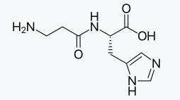
Additionally, when there is excessive sugar intake, excess glucose in the body can interact with proteins, amino acids, or lipids, leading to a process known as non-enzymatic glycation. This reaction results in the breakdown of collagen, leading to the loss of elasticity. Carnosine can act as a substitute, preventing glycation crosslinking of proteins, thereby helping to restore skin elasticity and brighten skin tone. Carnosine also helps maintain superoxide dismutase (SOD) activity, neutralizing the large amounts of lactic acid produced by muscles, and serving to buffer the physiological pH.
Carnosine is considered one of the earliest bioactive peptides used in the beauty and skincare industry, with nearly a century of chemical synthesis history and a well-established manufacturing process. It, along with copper peptides and palmitoyl tripeptides, is one of the most widely used peptides in cosmetics and is active in many international skincare brands. For instance, L’Oréal’s La Roche-Posay includes carnosine in several product lines, such as the La Roche-Posay Effaclar Duo+ Acne Treatment. Olay also incorporates carnosine in products like the Olay Regenerist Micro-Sculpting Serum. Additionally, brands like Chando and Elizabeth Arden also include carnosine in their formulations.
Carnosines at Creative Peptides
| CAT# | Product Name | M.W | Molecular Formula |
| CPC1634 | Carnosine | 226.23 | C9H14N4O3 |
| 10-101-223 | N-Acetyl Carnosine | 268.27 | C11H16N4O4 |
| CPC1681 | Decarboxy Carnosine HCl | 255.14 | C8H16Cl2N4O |
| R1914 | L-Homocarnosine | 240.26 | C10H16N4O3 |
Glutathione
Glutathione is a tripeptide compound containing a γ-amide bond and a thiol group. It is composed of glutamic acid, cysteine, and glycine and is present in nearly every cell of the body. The thiol group (-SH) on the cysteine residue is its active group. Under physiological conditions, glutathione mainly exists in its reduced form (G-SH), but it can also exist in an oxidized form (G-S-S-G). The two forms can interconvert through the action of the enzyme glutathione reductase.
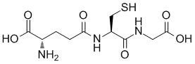
Recent studies have shown that glutathione can effectively reduce pigmentation by inhibiting the production of melanin, helping to prevent the formation of lipofuscin, or age spots. When used in combination with Vitamin C, it has been shown to have a good effect in treating melasma in women. Some experiments have also demonstrated that glutathione possesses certain tyrosinase inhibitory properties, making it a commonly used ingredient for whitening products.
Additionally, the thiol group (-SH) on the cysteine residue in glutathione exhibits strong nucleophilicity, allowing it to bind with heavy metal ions, drug metabolites, and other intermediates to form non-toxic or low-toxic complexes, which are then excreted from the body, playing a role in detoxification. Therefore, clinical treatments for melasma often combine glutathione with other agents such as the Jing Tian (Sedum) capsule for melasma removal, or with Vitamin C for treating dark spots.
Japanese brand Shiseido’s Anessa sunscreen is known for using a significant amount of glutathione. The famous French brand Filorga includes glutathione in its Brightening Collection, such as the Filorga Comfort Brightening Serum. In addition, brands like The Ordinary, Laneige, and Gu Yu also feature glutathione in their product formulations.
Frog Antimicrobial Peptide
Frog antimicrobial peptides are bioactive peptides extracted from the skin or secretions of amphibian species such as the Rana (tree frog). These peptides primarily come from the dorsal and ventral skin of the frog, serving as a defense mechanism developed through long-term natural selection. They typically consist of 10 to 50 amino acid residues and possess strong alkalinity, thermal stability, and broad-spectrum antimicrobial properties.
After the oil is extracted from the frog’s skin, it is rich in collagen and exhibits some degree of biocompatibility and degradability of functional proteins. It is considered a non-toxic active substance that, after self-assembly or cross-linking, can form collagen fibers with a certain strength and a degree of adhesion to wounds, offering a repair effect. The oil also contains a variety of amino acids and trace elements, including a high concentration of human chorionic gonadotropin, making it an ideal source of nutritional proteins. Theoretically, it also has antioxidant properties, and thus can be widely used in biotechnology and medical fields.
Compared to peptide-based substances, research and application of frog antimicrobial peptides are still in a relatively immature stage, with much of their potential yet to be developed. Current research focuses on enhancing the absorption and efficacy of frog antimicrobial peptides through biotechnology. For example, materials like chitosan are being used to prepare composite sponges that improve adhesion and water absorption, making them suitable raw materials for developing reparative skincare products.
Other peptides (Plant-Derived)
Soy protein-derived antioxidant peptides are a class of plant-derived peptides and an important branch of natural antioxidant peptides. These peptides are typically extracted from crops and vegetables through protein hydrolysis, and include varieties such as wheat bran peptides, mung bean peptides, soy peptides, and tea seed peptides. Among these, soy peptides have the longest history of development and use. They not only address issues like the poor solubility and instability of soybean proteins but also, through ultrafiltration enzymatic hydrolysis, produce peptide fragments with high antioxidant activity. These peptides are especially effective in inhibiting the formation of singlet nitrogen species.
The ability of soy peptides to scavenge hydroxyl radicals is closely related to the exposure of their amino acid side chains. Their antioxidant mechanisms are diverse and include, but are not limited to, directly capturing reactive oxygen species (ROS), activating the body’s antioxidant enzyme system, and chelating metal ions, thereby exerting a comprehensive antioxidant effect. These properties make soy protein-derived antioxidant peptides widely applicable in nutritional supplements, functional foods, and cosmetics.
Comparison of efficacy
| Type of Peptides | Functional Characteristics |
| Copper Peptide | Promotes the production of collagen and glycosaminoglycans, helping to enhance wound healing and skin tightening.Has antioxidant properties, reducing damage to the skin caused by free radicals.Possesses anti-inflammatory effects, aiding in skin inflammation repair.Helps improve signs of skin aging and enhances skin elasticity. |
| Carnosine | Possesses strong antioxidant capacity, clearing free radicals in the body and protecting cells from oxidative damage.Helps reduce melanin production, improving skin tone.Can reduce wrinkles and increase skin hydration.Contributes to maintaining immune system stability and cell viability.In athletics, enhances physical strength and endurance. |
| Glutathione | Acts as an internal antioxidant weapon of the body, providing widespread antioxidant effects and protecting cells from oxidative damage.Has detoxifying, whitening, and anti-aging physiological effects.Assists in removing toxic substances and repairing liver damage.Improves immunity and other functions.When used in skin care, helps whiten skin and improve uneven skin tone. |
| Frog Antimicrobial Peptide | Has immune-boosting properties, inhibiting the activity of viruses, bacteria, and tumors.Strengthens the body’s immune system, enhancing resistance.In clinical use, shows antiviral, anticancer, and liver-protecting effects. Examples include hepatitis treatment.As an immune factor, thymosin peptide is widely used in antiviral and health-support therapies. |
What is solid-phase peptide synthesis (SPPS)?
Solid-phase peptide synthesis (SPPS) is an important method for synthesizing peptides. In SPPS, peptides are synthesized on an insoluble resin that serves as the solid support. The process begins by attaching the C-terminal amino acid to the resin, and subsequent amino acids are added step by step in a sequence dictated by the desired peptide. Each elongation cycle typically consists of deprotection (removing a protecting group from the amino acid) and condensation (forming a peptide bond between the activated amino acid and the growing chain).
The most challenging part of peptide synthesis is the formation of the peptide bond, which requires an efficient activation of the carboxyl group to promote its reaction with the amino group. This activation is achieved through the use of condensation agents, which are chosen based on factors such as compatibility with the resin, reaction efficiency, and minimization of side reactions.
General process of synthesis
- First, the initial amino acid (the C-terminal residue) is attached to the resin (loading).
- After the amino acid binds to the resin, the resin is washed to remove by-products and excess reagents.
- Next, the amino protecting group is removed, and the resin is washed again. Under the action of a coupling agent, the next amino acid is coupled to the peptide-resin complex. This coupling and washing cycle is repeated until the entire peptide sequence is completed.
- Finally, all protecting groups are removed, the peptide-resin is washed, and the peptide is cleaved from the resin.
Key Considerations in Choosing a Condensation Agent
The selection of a condensation agent is crucial for ensuring efficient peptide bond formation. Key factors influencing the choice of a condensation agent include:
Activation Efficiency: The reagent must activate the carboxyl group of the amino acid efficiently to form the reactive intermediate, which will then react with the amine group of the next amino acid.
Minimization of Side Reactions: The condensation agent should ideally minimize side reactions such as racemization, hydrolysis, and degradation of sensitive amino acids or functional groups.
Compatibility with the Resin: The reagent must not interfere with the resin or other components of the reaction mixture, including solvents and protecting groups.
Solubility: Condensation agents should be sufficiently soluble in the solvent system used during SPPS to achieve optimal reaction conditions.
“Orthogonal” protection in solid-phase peptide synthesis
Many amino acid side chains are reactive and, if not protected, can form by-products. To successfully synthesize peptides, side chains need to be protected, and these protecting groups must remain stable throughout repeated deprotection of the amino group. Ideally, the amino-protecting group and side-chain-protecting group can be removed under completely different conditions-such as removing the amino-protecting group under basic conditions and the side-chain-protecting group under acidic conditions. This approach is known as “orthogonal” protection. Common combinations include Boc/Bzl protection and Fmoc/tBu protection. In the Boc/Bzl protection scheme, the Boc group temporarily protects the amino group, while the benzyl group provides more permanent protection for side chains. Although both Boc and benzyl protecting groups are acid-sensitive, making Boc/Bzl not a truly orthogonal protection strategy, it is still widely used. This is because the Boc group can be removed under mild acidic conditions (e.g., 50% TFA in DCM solution), whereas the benzyl group requires a very strong acid (such as HF or TFMSA) for removal.
Condensing agents in solid-phase synthesis
Carbodiimides
Carbodiimides, such as dicyclohexylcarbodiimide (DCC) and 1-(3-dimethylaminopropyl)-3-ethylcarbodiimide (EDC), are among the most widely used condensation agents in peptide synthesis. DCC, DIC, EDCI. Dicyclohexylcarbodiimide (DCC) and diisopropylcarbodiimide (DIC) are commonly used to convert carboxylic acids into amides, esters, and anhydrides. They can also convert primary amides into nitriles, which may lead to side reactions when aspartic acid and glutamic acid residues are present in peptides. DCC generates dicyclohexylurea as a by-product, which is nearly insoluble in most organic solvents and precipitates out of the reaction mixture as the reaction progresses. Therefore, DCC is highly useful in solution-phase reactions but less suitable for reactions on resin. In solid-phase synthesis, DIC is used because its urea by-product is more soluble and remains in solution. In some applications, such as protein modification, ethyl-(N’,N’-dimethylaminopropyl) carbodiimide hydrochloride (EDCI) is used. This carbodiimide reagent and its urea by-product are water-soluble, allowing by-products and excess reagent to be removed with water washes.
In peptide synthesis, to prevent racemization of amino acids during carbodiimide activation, an equivalent amount of 1-hydroxybenzotriazole (HOBt) is commonly added along with the carbodiimide.
Mechanism of Action: Carbodiimides activate the carboxyl group of an amino acid by forming an acyl-urea intermediate. This intermediate then reacts with the amino group of another amino acid, resulting in the formation of a peptide bond.
Advantages: Carbodiimides are relatively inexpensive and easy to handle. They can be used for a wide variety of amino acids and peptides, and they are effective under mild conditions, minimizing the risk of side reactions such as racemization.
Limitations: One of the significant drawbacks of carbodiimides is the formation of urea byproducts, such as dicyclohexylurea (DCU) when DCC is used. These byproducts can precipitate out of solution and require removal by filtration, which can reduce the yield. Moreover, carbodiimides can also lead to the formation of side products due to incomplete activation.
Benzotriazole Derivatives
Benzotriazole derivatives, such as 1-hydroxybenzotriazole (HOBt), benzotriazole-1-yl-oxy-tris-pyrrolidino-phosphonium hexafluorophosphate (BOP), and Oxyma, are frequently used in peptide synthesis to improve the efficiency of carbodiimide-based activation.
To avoid racemization and side reactions associated with carbodiimide condensing agents, many alternative reagents have been developed to generate OBt esters in situ. Benzotriazol-1-yloxytris(dimethylamino)phosphonium hexafluorophosphate (BOP) was one of the first reagents developed. BOP does not lead to dehydration of asparagine and glutamine side-chain amides to form nitrile by-products, and it causes minimal racemization of amino acids. However, BOP produces highly carcinogenic hexamethylphosphoramide as a by-product in condensation reactions, so it must be handled carefully. PyBOP offers comparable condensation efficiency to BOP but with a lower risk from its by-products. PyAOP is also an effective condensing agent, especially for coupling between N-methyl-protected amino acids. PyBrOP is often used for coupling sterically hindered amino acids, such as N-methyl amino acids or α,α-dialkylglycines, where other condensing agents are less efficient. BOP-Cl is commonly used to couple Fmoc-α,α-dialkyl amino acids to tritylphenyl resins.
Mechanism of Action: These reagents work by stabilizing the active ester intermediate that is formed during the activation process. HOBt, for example, reacts with the carbodiimide to produce a highly reactive ester intermediate, enhancing the efficiency and reducing side reactions.
Advantages: The addition of HOBt or related compounds can significantly improve the efficiency of peptide bond formation, reduce side reactions, and suppress racemization, especially for sterically hindered or sensitive amino acids.
Limitations: While these reagents are effective, their cost can be higher compared to carbodiimides alone, and their use requires additional care in handling and disposal due to their chemical reactivity.
Uronium Salts
Uronium salts, such as ethyl(2-dimethylamino-ethyl)-phosphonium tetramethylfluorophosphate (EEDQ) and t-butylphosphonium hexafluorophosphate (TBTU), are another class of reagents used in peptide bond formation. These agents activate carboxyl groups by forming reactive phosphonium or uronium species.
Mechanism of Action: Uronium salts generate reactive species that facilitate the coupling of the carboxyl group with the amine group of the incoming amino acid. These species are highly electrophilic, promoting efficient peptide bond formation.
Advantages: Uronium salts are highly effective in promoting fast and efficient peptide bond formation. They are particularly useful for synthesizing peptides with low solubility or challenging sequences. They also tend to produce fewer side products.
Limitations: Despite their efficiency, uronium salts can be expensive and may lead to contamination with byproducts if not carefully controlled. Their use in SPPS may also be limited by the compatibility with the resin or other reagents.
Phosgene Derivatives
Phosgene derivatives, such as chlorophosgene and triphosgene, are potent agents used for activating carboxyl groups in peptide synthesis.
Mechanism of Action: Phosgene derivatives react with the carboxyl group of amino acids to form an acylated intermediate. This intermediate then reacts with the amine group of the next amino acid, facilitating the peptide bond formation.
Advantages: Phosgene derivatives are highly efficient in peptide bond formation and can work well with challenging amino acids. They offer high yields and can help minimize racemization and side reactions.
Limitations: The major disadvantage of phosgene derivatives is their toxicity and safety concerns, as phosgene is a highly hazardous substance. Additionally, the reagents can be expensive, and their use requires specialized equipment for handling and disposal.
Activation with Mixed Anhydrides
Mixed anhydrides are formed by reacting an amino acid with an acid chloride or an acylating agent. This reaction produces an acylated amino acid intermediate that is then used for peptide bond formation.
Mechanism of Action: The mixed anhydride is a reactive intermediate that can readily react with the amino group of another amino acid, resulting in the formation of a peptide bond.
Advantages: Mixed anhydrides can be highly effective, especially when used in combination with reagents like HOBt or pyridine. The method is versatile and applicable to a broad range of amino acids.
Limitations: One challenge with this approach is that mixed anhydrides are highly reactive, which can sometimes lead to side reactions if the reaction conditions are not carefully controlled. Additionally, the preparation of mixed anhydrides can be more complex than other methods.
New Developments in Condensation Agents
Recent advances in peptide synthesis have led to the development of newer, more efficient condensation agents. These agents aim to address the limitations of traditional reagents by enhancing reaction specificity, improving yields, and reducing side reactions.
One promising area is the development of green reagents, which are designed to be environmentally friendly and non-toxic. For example, Oxyma Pure, a derivative of HOBt, is one such reagent that has gained popularity due to its improved efficiency and lower toxicity profile.
Additionally, solid-phase activated reagents (SPARs) are being explored as a way to streamline the synthesis process by coupling the activation step directly to the solid-phase resin. These reagents eliminate the need for liquid-phase activation, which can reduce solvent consumption and simplify the overall process.
]]>Trofinetide is a synthetic glycine-proline-glutamic acid (GPE) tripeptide analogue derived from the N-terminal sequence of insulin-like growth factor 1 (IGF-1). It is primarily used to treat Rett syndrome (RTT), a rare inherited neurodevelopmental disorder that primarily affects women and is usually caused by mutations in the MECP2 gene.
Trofinetide works by reducing neuroinflammation and supporting synaptic function. Its mechanism of action includes enhancing synaptic activity, restoring synaptic structure, inhibiting the effects of inflammatory compounds in the brain, increasing antioxidant responses, reducing injury-induced apoptosis, and increasing the presence of IGF-1 in the central nervous system. In addition, Trofinetide has also been found to be able to inhibit the production of inflammatory cytokines and inhibit the overactivation of microglia and astrocytes.
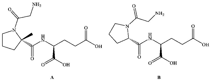
Trofinetide is the first FDA-approved drug for the treatment of Rett syndrome in adults and children 2 years of age and older. In clinical trials, Trofinetide has shown significant improvements in the core symptoms of Rett syndrome, such as communication, behavior, and motor function. However, although its efficacy has been validated, its exact mechanism of action needs further research. Trofinetide is an innovative therapy for Rett syndrome that improves the neurological function and structure of patients through multiple mechanisms, bringing new hope to this disease that has long lacked effective treatments.
Trofinetide in Rett Syndrome
Rett syndrome is a rare genetic disorder that affects psychomotor development in children, mainly due to mutations in the methyl-binding protein 2 (MECP2) gene on the X chromosome. Rett syndrome is a neurodevelopmental disorder that affects female children and is characterized by loss of verbal communication, stereotyped hand movements, seizures, and behavioral problems. Trofinetide, through its synthetic glycine-proline-glutamate analogue properties, aims to improve the adverse pharmacokinetic profile of naturally occurring GPE in the brain, thereby positively affecting the symptoms of patients with Rett syndrome.

How does Trofinetide Work?
Rett syndrome is a rare inherited neurodevelopmental disorder whose core pathological mechanism is closely related to mutations in the MECP2 gene. The MECP2 gene plays an important role in brain development and is involved in regulating gene expression, synaptic function, and homeostasis of the nervous system. However, when the MECP2 gene is mutated, these functions are significantly weakened or lost, leading to synaptic abnormalities, increased neuroinflammation, and degeneration of nerve cells. These changes are the main cause of core symptoms such as speech impairment, motor coordination difficulties, and cognitive deterioration in patients with Rett syndrome.
As an innovative therapy for Rett syndrome, Trofinetide’s mechanism of action precisely targets the above-mentioned pathological processes, aiming to comprehensively improve the neurological function and symptomatic manifestations of patients. The following are the specific mechanisms by which Trofinetide works at multiple levels:
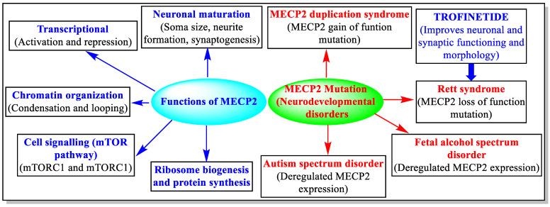
Improves Synaptic Function
Synapses are key sites for signal transmission between neurons, and their structural and functional integrity directly affect the normal functioning of brain function. In patients with Rett syndrome, synaptic plasticity is reduced and nerve signaling is inhibited due to defects in the MECP2 gene, ultimately leading to significant deterioration of cognitive and behavioral abilities. Trofinetide is able to promote normal signaling between neurons by enhancing synaptic plasticity. In addition, Trofinetide repairs damaged synaptic structures, radically improving the brain’s signaling network. This effect not only helps to restore the patient’s cognitive abilities, but also improves their language and social interaction skills.
Inhibits Inflammatory Responses
Neuroinflammation is one of the important pathological features of Rett syndrome, which is characterized by excessive release of inflammatory cytokines and abnormal activation of glial cells. The inflammatory response can cause further damage to neurons and exacerbate the neurological deterioration of the patient. Trofinetide is able to inhibit the inflammatory response through a variety of mechanisms. On the one hand, it can reduce the release of inflammatory cytokines such as IL-6 and TNF-α, thereby reducing the damage of inflammation to neurons. On the other hand, it is able to inhibit the overactivation of astrocytes and microglia and maintain the stability of the nervous system microenvironment. This effect helps to alleviate the neurodegenerative changes of the patient and creates favorable conditions for the restoration of neurological function.
Promotes Antioxidant Reactions
Oxidative stress is another significant pathological feature of Rett syndrome. Due to the excessive accumulation of free radicals, the patient’s nerve cells face severe oxidative damage, which can further weaken brain function. Trofinetide has a powerful antioxidant capacity, which can reduce the production of free radicals and promote the activity of antioxidant enzymes, thereby reducing the damage of oxidative stress to nerve cells. This antioxidant effect not only protects the structural integrity of nerve cells, but also slows the disease progression and provides patients with more stable support for nervous system function.
Cell Protection and Regeneration
The brains of patients with Rett syndrome are often accompanied by increased apoptosis and decreased regenerative capacity of nerve cells. Trofinetide is able to protect nerve cells from damage through a variety of mechanisms while promoting their regeneration. On the one hand, it can reduce the apoptosis of neurons and prolong the survival time of cells. On the other hand, it is able to increase levels of insulin-like growth factor 1. IGF-1 plays a key role in promoting neuronal growth and repair, providing regenerative support to damaged nerve cells, thereby enhancing the patient’s brain repair capacity. This cytoprotective and regenerative effect is the core foundation of Trofinetide’s improvement of Rett syndrome symptoms.
Comprehensive Multi-dimensional Improvement
Through the above mechanisms, Trofinetide can not only improve the core symptoms of patients with Rett syndrome, but also improve the functional stability of the nervous system as a whole. This multidimensional effect makes trofinetide a very promising treatment option, bringing hope to patients with Rett syndrome who have long lacked effective treatments.
Development of Trofinetide
The U.S. Food and Drug Administration (FDA) approved Trofinetide (brand name Daybue) in March 2023 for the treatment of Rett syndrome in adults and children 2 years of age and older. The approval is based on a Phase 3 clinical trial called Lavender, which evaluated the efficacy and safety of trofinetide in 187 women aged 5 to 20 years with Rett syndrome.
In a 12-week randomized, double-blind, placebo-controlled trial, the study evaluated the efficacy and safety of trofinetide in patients with Rett syndrome. The primary endpoints of the trial included change in the Rett Syndrome Behavior Questionnaire (RSBQ) total score, which assesses improvement in core behavioral symptoms, and the Clinical Global Impression of Improvement Scale (CGI-I) score, which measures change in the patient’s overall condition. The results showed that the Trofinetide group demonstrated a significant advantage over placebo in both primary endpoints, with significant improvements in communication, attention, and adaptive behavior, as well as a significantly higher proportion of physicians rating patients for overall symptom improvement, further confirming the clinical efficacy of Trofinetide.
Summary
Trofinetide marks an important breakthrough in this area as the first FDA-approved treatment specific to Rett syndrome. Its unique mechanism of action, multi-dimensional neuroprotective function and remarkable efficacy in clinical trials provide new hope for the treatment of patients with Rett syndrome.
While the approval of Trofinetide is a milestone, there are still areas to explore.
Long-term Efficacy and Safety: Larger, long-term studies are needed to validate the ongoing efficacy and potential side effects of trofinetide.
Individualized Treatment: To further study the differences in patients’ responses to Trofinetide to provide the best treatment options for patients with different genotypes or symptom characteristics.
Combination Therapy Possibilities: The effects of trofinetide in combination with other potential drugs or treatments deserve further discussion.
The advent of Trofinetide has ushered in a new era in the treatment of Rett syndrome and provided valuable experience for drug development for other rare neurodevelopmental disorders.
References
- Parent, Harrison, et al., Trofinetide: a pioneering treatment for Rett syndrome. Trends in Pharmacological Sciences (2023).
- Hudu, Shuaibu A., et al., Trofinetide for Rett syndrome: highlights on the development and related inventions of the first USFDA-approved treatment for rare pediatric unmet medical need. Journal of Clinical Medicine 12.15 (2023): 5114.
As early as the 80s of the last century (1983), the exploration of oral delivery of peptides has achieved a historic breakthrough through cyclosporine A (Fig.1). This natural peptide can achieve high oral bioavailability and is therefore developed as an oral formulation.
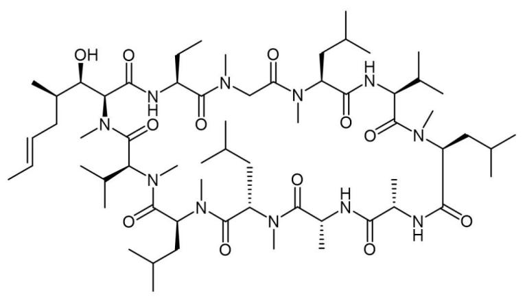
However, the oral delivery of Cyclosporine A has stimulated people’s enthusiasm for the development of oral peptides, but the key to completely opening the door to oral peptides has not been fully presented, and people are still searching for the realization of oral bioavailability of peptides. In addition to the stability of peptides and their resistance to degradation by a variety of enzymes, how to cross the cell membrane is also one of the focuses of research. Researchers from the University of ETH in Switzerland recently simulated the molecular dynamics of peptide crossing the cell membrane using a flexible decacyclic peptide (Fig.2).
In contrast to straight-chain peptides, which are too flexible, cyclic peptides have what are known as chameleon properties. Chameleons change skin color based on the environment, while cyclic peptides can change the molecular conformation based on the polarity of the environment. This property depends on the conversion of intramolecular and intermolecular hydrogen bonds in the cyclic peptide molecule.
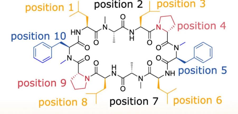
In the so-called Open conformation of investigational peptide drugs, the hydrogen-bonded atoms of cyclic peptides are exposed to a polar environment (e.g., in blood or cytoplasm) to form hydrogen bonds with their counterparts in solvents, or with polar solvent molecules. But when the environment becomes non-polar (e.g., inside a cell membrane), cyclic peptides can instead adopt the so-called Closed conformation, wrapping themselves up and encapsulating themselves by forming intramolecular hydrogen bonds, encapsulating polar groups inside the molecule and exposing the non-polar structure to surface of the molecule, thereby reducing the polar area and the desolvation energy (the amount of energy required for the solute to be removed from the water or other solvents). This chameleon property of cyclic peptide is helpful in the development of oral peptide formulations because it can balance good water and fat solubility, which favors increasing cell permeability.
The Closed state is generally considered to be the predominant permeability state of cyclic peptides. However, it is a dynamic process that is controlled by a variety of factors such as sequence, molecular size, hydrophobic surface area, etc. Through molecular dynamics simulation, the researchers simulated the process of cyclic peptide molecules crossing the cell membrane through passive osmosis into the cell, highlighting the importance of flexible conformation for cyclic peptide membrane permeation, and dividing it into four steps (Fig.3):
- Specific peptide side-chain residues can act as molecular anchors to establish the initial contact of cyclic peptides with the cell membrane before the peptide molecule is inserted into the cell membrane.
- After anchoring to the cell membrane, the peptide molecule jumps directly into it, inserting itself into the interface between the polar head and non-polar tail regions of the cell membrane. In this process, the cyclic peptide exhibits two potential directions of arrangement, and the open state takes orientation A and B, and preference orientation A is preferred. Closed cyclic peptides take orientation B only.
- The Open state transitions to the Closed state, increasing the chance of it penetrating. At this time, the chameleon nature of the peptide molecules is very important for them to move from a polar environment to a non-polar environment. It is difficult for the cyclic peptide in the open state to achieve the subsequent diffusion process.
- Only cyclic peptide molecules in the closed state are likely to diffuse from this polar/non-polar mixed environment on the cell membrane surface through the non-polar lipid membrane leaflet and spread to the polar/non-polar interface on the other side. Through the anchoring and flipping mechanism, the final transmembrane journey is achieved.
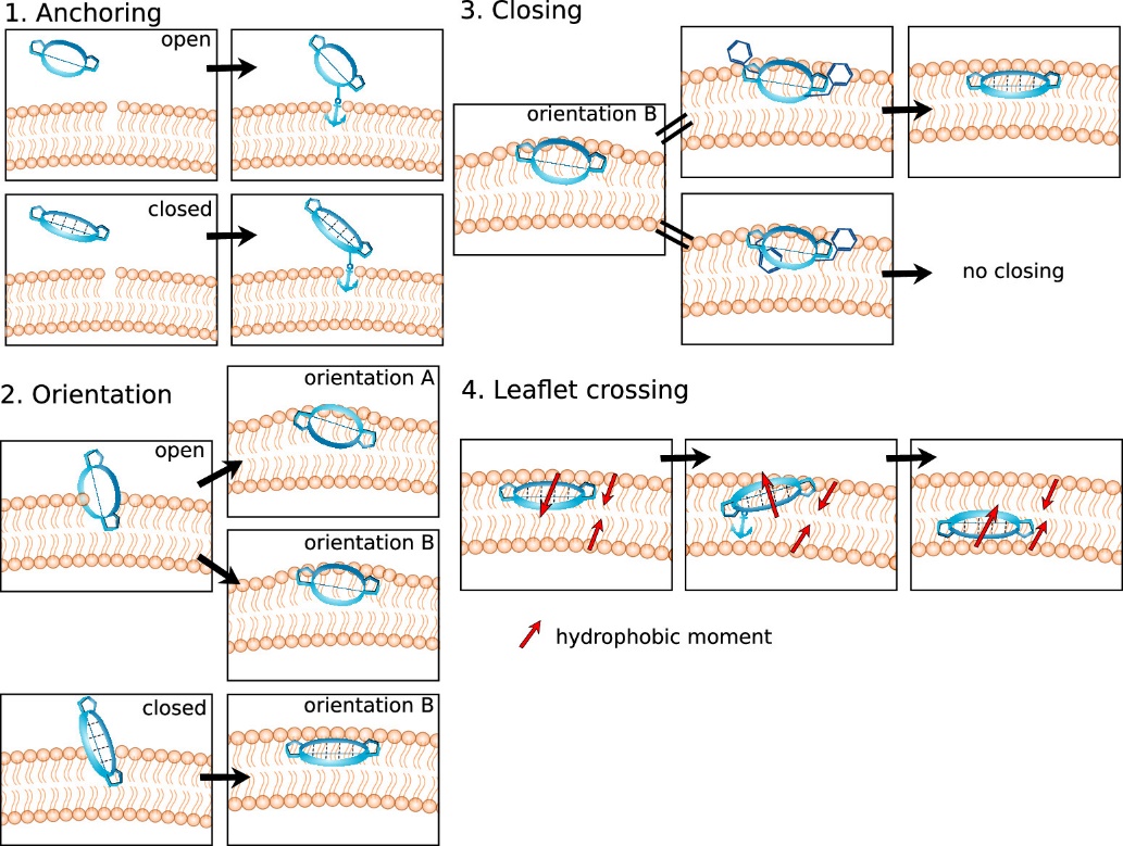
Step 1
Simulated molecular dynamics showed that the anchor residues of the Open conformation and the Closed conformational cyclic peptide were different, due to the fact that the degree of contact of the residues was not the same in the two conformations. Given that the anchoring of amino acids is peptide conformation-dependent, characterizing the conformational behavior of cyclic peptides is critical for rational peptide drug design. Understanding the main conformations of peptides before they enter the cell membrane and the corresponding residue exposure will help optimize the design of membrane-permeable cyclic peptides. This mode of membrane permeability initiated by side chain anchoring of amino acid residues can be generalized to other molecular modalities other than peptides, such as increasing the membrane permeability of molecules by means of moderate aliphatic chain conjugation.
Step 2
The peptide is inserted into the membrane and parallels itself to the membrane plane. If the insertion occurs in a molecule with an open conformation, the peptide may take two directions of arrangement, A and B. However, if the peptide insertion is already in a closed conformation (pre-folded), only direction B is observed. This can also be useful for the design of other macrocyclic peptides, especially those with continuous hydrophobic surfaces. Only orientation B favors the conversion of peptides from the Open to Closed conformation.
Step 3
The change of conformation from the open state to the closed state is a critical process that has an important impact on the subsequent intramembrane diffusion. This conformational transition not only changes the spatial structure of the molecule, but also significantly affects its interaction with the membrane environment, thus determining the movement mode and diffusion efficiency of the molecule in the membrane.
Step 4
The peptide molecule that enters the cell membrane must expand to the other side of the membrane (lower leaflet). As the final step of crossing the membrane, the peptide molecule needs to go through a similar process as the first step, that is, its amino acid residues need to be anchored to the polar head of the lower leaflet, and this process also involves the reversal of the direction of the cyclic peptide, resulting in a change in the direction of the hydrophobic moment.
Simulations have shown that the effects of amino acids (e.g., leucine, phenylalanine, proline) may be highly correlated with the peptide sequence, depending on the position of the amino acid in the peptide as well as other residues. The goal of this molecular dynamics simulation of the cyclic peptide permeation pathway is to improve the bioavailability of peptides, especially oral bioavailability, and to develop more peptide drugs that can be delivered orally.
References
Linker, Stephanie M., et al., Lessons for Oral bioavailability: how Conformationally flexible cyclic peptides enter and cross lipid membranes. Journal of medicinal chemistry 66.4 (2023): 2773-2788.
]]>Non-ribosomal peptide synthesis is a universal and critical biochemical process catalyzed by NRPSs in bacteria and fungi, through which a wide array of therapeutically important peptides with highly diverse structures and bioactivities, including penicillin, bleomycin, and cyclosporine, are produced. Biocatalysts capable of NRP synthesis could be divided into two groups (ATP-independent and ATP-dependent enzymes) based on the difference in substrate activation. In the ATP-dependent process, enzymes such as tRNA-dependent ligase can activate the substrate through aminoacyl-adenosine monophosphate. In the ATP-independent process, enzymes such as transacylase use aminoacyl phosphate.
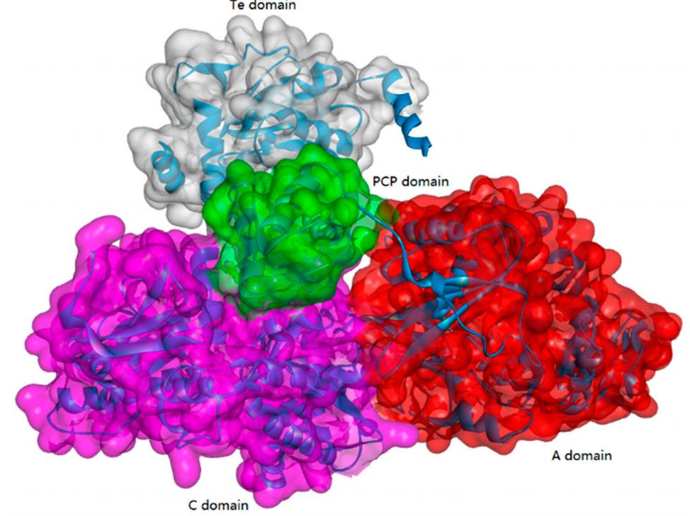

Peptide Synthesis at Creative Peptides
| Services | Description |
| Peptide Synthesis Services | We provide high-quality customized peptide synthesis services, from bulk API peptides, high-throughput library peptides, cosmetic peptides, to array peptides, antigen peptides, and other complex or abnormal peptide sequences. |
| Peptide Modification Services | Creative Peptides offers hundreds of peptide modifications to meet any research need. These modifications can improve the overall stability of peptide, change the structure to better understand biological functions, or enhance the immunogenicity of antibody development and production. |
| Custom Conjugation Service | Creative Peptides provides convenient multiple types of peptide conjugation services to meet the needs of different projects, including activating immune responses, obtaining higher stability and cell penetration. |
ATP-Grasp Enzymes
The ATP-grasp enzymes, also known as ATP-dependent carboxylate-amine ligases, activate acids like acylphosphate intermediates and are found in various biological systems such as de novo purine biosynthesis. Notable members of this family include biotin carboxylase, Ddl, and glutathione synthetase. ATP-grasp enzymes usually have a unique structure with three conserved domains, with a nonclassical ATP binding fold enclosing an ATP molecule. Most of these enzymes require an Mg2+ ion coordinated by ATP in the active site.
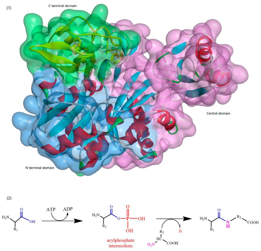
α-Amino Acid Ester Acyltransferase
Kenzo and colleagues reported an efficient enzymatic method for producing oligopeptides from unprotected amino acids with high yield, using Empedobacter brevis ATCC 14234 strain. An enzyme catalyst (named carboxypeptidase Y) was found in the strain to assist in rapid production of certain oligopeptides. However, details about the amino acid sequence, coding gene sequence or 3D crystal structure were not provided. Isao ABE and team first reported about the cloning and expression of α-amino ester acyl transferases (AETs) from the same and another strain, their amino acid sequences were 35% and 36% identical to the α-amino acid ester hydrolase from Acetobacter pasteurianus. AETs displayed dual action-dipeptidyl peptidase and transferase activities, and was quite specific for both acyl donors and nucleophiles. There have been no studies yet on the 3D structure of AET or its reaction mechanisms.

Enzymes Used for β-Lactam Biosynthesis (β-Lactam Acylases)
β-Lactam antibiotics, which contain a β-lactam ring in their chemical structure and include types such as penicillin, cephalosporins, and thiamycins, are widely used as anti-infective agents and have significant importance in the pharmaceutical industry. The environmentally-friendly and cost-effective synthesis of β-lactam antibiotics via enzymatic means has been increasingly applied in modern pharmaceutical enterprises. β-lactam acylases, which historically process β-lactam antibiotics, can also be used for the creation of semi-synthetic β-lactam antibiotics. Different types of β-lactam acylases such as penicillin acylase (PA), glutaryl acylase (GA), and β-amino acid ester hydrolase (AEH) have been studied in the biosynthesis of β-lactam antibiotics. PAs are generally produced by a variety of microorganisms and are categorized into two types based on their substrate specificity. They are widely used in the industrial production of the active pharmaceutical intermediate 6-aminopenicillanic acid (6-APA) and for the synthesis of semi-synthetic antibiotics, making them potentially useful in the development of novel drugs. Furthermore, PAs can be used in peptide synthesis, resolution of racemic mixtures, and in the production of both achiral and chiral compounds for pharmaceutical intermediates.
Cyanophycinases (CGPases)
Cyanophycin granule polypeptide (CGP, or multi-L-arginyl-poly) is an intracellular storage polymer found in most cyanobacteria. Equimolar concentrations of arginine and aspartic acid are observed in the aspartic acid backbone, where the arginine moieties are linked to the β-carboxyl group of each aspartic acid through its α-amino group. In most genera of cyanobacteria, the cyanophycin synthetase gene (cphA) has been identified and verified for the synthesis of CGP. In contrast, the intracellular and extracellular degradation of CGP is catalyzed by cyanophycinases (CphB and CphE), which releases dipeptides (β-Asp-Arg). In this respect, β-Asp-Arg can be efficiently synthesized via the simultaneous production of CGP and CGPase, which could be further applied in various fields requiring arginine (Arg) content in feed or food. However, the production and efficient isolation of CGP from various organisms have been successfully established in several recombinant strains, including Escherichia coli, Nicotiana tabacum, Pseudomonas putida, and Pseudomonas alcaligenes DIP1. Therefore, it is very feasible to produce dipeptides (e.g., β-Asp-Arg) via the metabolic engineering of suitable hosts and chemo-enzymatic strategies. For example, successful co-expression of CGP and CGPase in the Nicotiana tabacum plant was recently achieved A further study showed that it is possible to realize the goal of sufficient storage and efficient transport of arginine and β-Asp-Arg dipeptides in this synthetic model.
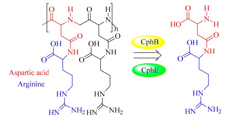
Reference:
- Wang, T., et al. Strategy for the biosynthesis of short oligopeptides: Green and sustainable chemistry. Biomolecules. 2019, 9(11): 733.