Fluorescent techniques have revolutionized biochemical research over the last 20 years. Because they have characteristics of high sensitivity, selectivity, fast response time, flexibility and experimental simplicity, the use of fluorescent techniques in biological research is very widespread. In many cases, biological sensors consist of fluorescent versions of a particular protein.
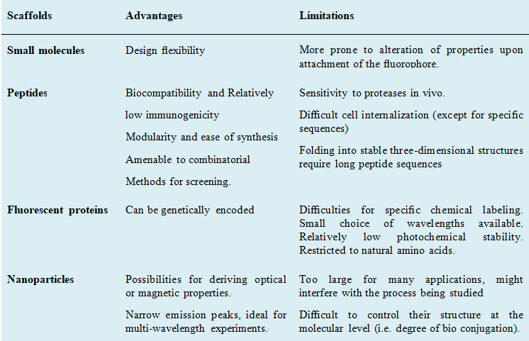
 Fig. 1 Advantages and limitations of different fluorescent probes.
There are different distances between different fluorophores that affect the fluorescent signal. An interesting strategy for the synthesis of sensors involves the application to peptides of the molecular beacon concept that was developed in the field of nucleic acid sensors. The development of peptide-based sensors over the last two decades has been spectacular: FRET effects or environment sensitive fluorophores provide reliable design strategies that can be safely implemented to study virtually any biological interaction with minimal effort. The large number of commercially available fluorophores, many of them in the form of activated species ready for derivatization, has simplified the synthetic effort required to obtain the modified peptides and increased the flexibility of the designs. Given the increasing interest in the development of chemical tools for biological research, the importance of these peptide probes is only expected to grow. Creative Peptides provides available fluorescence and dye labeling peptides services to meet your custom peptide need.
Fluorescence labeling of peptides involves the conjugation of peptides with fluorescent dyes or probes, enabling visualization and tracking of peptide localization, behavior, and interactions within biological systems. This process harnesses the unique optical properties of fluorescent molecules, which emit light of specific wavelengths upon excitation by an external light source.
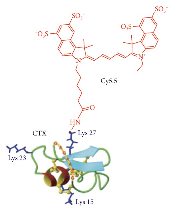
 Fig. 2 Peptide chlorotoxin (CTX) is labeled with Cy5.5. (Joshi, B. P., 2018)
Fluorescent-labeled peptides find utility across various disciplines, including basic research, drug discovery, and clinical diagnostics. They are particularly advantageous in studies requiring real-time monitoring of cellular processes, such as receptor-ligand binding, protein-protein interactions, and intracellular trafficking. Additionally, they serve as valuable tools for fluorescence microscopy, flow cytometry, and high-throughput screening assays.
Visualization: Fluorescent labels allow direct visualization of the peptide within cells or tissues using fluorescence microscopy or other imaging techniques. This aids in studying peptide localization, trafficking, and interactions within biological systems.
Quantification: Fluorescence intensity can be quantitatively measured, enabling accurate determination of peptide concentrations in biological samples. This is particularly useful in pharmacokinetic studies and quantitative assays.
Multiplexing: Different fluorescent labels can be attached to peptides, enabling multiplexing and simultaneous detection of multiple peptides or targets within the same sample. This is valuable for studying complex biological processes involving multiple molecules.
High sensitivity: Fluorescent detection methods typically offer high sensitivity, allowing the detection of low concentrations of labeled peptides. This is advantageous for applications requiring high sensitivity, such as detecting biomarkers or studying low-abundance molecules.
Real-time monitoring: Fluorescent-labeled peptides can be used for real-time monitoring of biological processes, such as enzyme activity, receptor binding, or signal transduction events. This dynamic monitoring provides insights into the kinetics and dynamics of biological processes.
Versatility: Fluorescent labels come in a variety of colors and chemical properties, offering versatility in experimental design. Researchers can choose labels with specific excitation and emission wavelengths to minimize background fluorescence and spectral overlap, enhancing signal specificity.
Compatibility: Fluorescent labeling techniques are compatible with a wide range of peptide sequences and structures, allowing labeling of peptides without significantly altering their biological activity or properties.
Ease of detection: Fluorescent detection methods are relatively simple and accessible compared to other labeling techniques such as radioactive or mass spectrometry-based methods. This simplifies experimental procedures and reduces the need for specialized equipment.
Cellular imaging: Fluorescent-labeled peptides are frequently used as probes for imaging specific cellular structures or processes. They can target specific proteins, organelles, or cellular compartments, allowing researchers to visualize them under a microscope. This is crucial for understanding cellular dynamics and studying disease mechanisms.
Drug delivery: Peptides labeled with fluorescent dyes can be used to track the delivery of drugs or therapeutic agents in living organisms. By attaching fluorescent tags to peptides, researchers can monitor the distribution and uptake of drugs in real-time, helping to optimize drug delivery strategies and improve treatment efficacy.
Protein-protein interaction studies: Fluorescent-labeled peptides can be used to study protein-protein interactions in vitro and in vivo. By labeling peptides with different fluorophores, researchers can monitor the binding of proteins to each other, providing insights into complex biological pathways and signaling networks.
Biosensing: Fluorescent-labeled peptides can serve as sensitive biosensors for detecting specific molecules or analytes. By designing peptides that undergo a conformational change or fluorescence resonance energy transfer (FRET) in the presence of a target molecule, researchers can develop highly selective and sensitive detection methods for applications such as medical diagnostics and environmental monitoring.
High-throughput screening: Fluorescent-labeled peptides are valuable tools for high-throughput screening assays, allowing researchers to rapidly screen large libraries of compounds for potential drug candidates or bioactive molecules. By monitoring changes in fluorescence intensity or localization, researchers can identify compounds that modulate specific biological processes or protein targets.
Labeling and tracking biomolecules: Fluorescent-labeled peptides can be used to label and track biomolecules such as proteins, nucleic acids, and carbohydrates. This is useful for studying the dynamics of biomolecular interactions, trafficking, and localization within cells and tissues.
Materials science: Fluorescent-labeled peptides can be incorporated into various materials, such as hydrogels, nanoparticles, and polymers, for applications in biotechnology and materials science. By engineering peptides with specific functional properties and incorporating fluorescent tags, researchers can develop advanced materials for drug delivery, tissue engineering, and sensing applications.
Cell penetration monitoring: Fluorescent-labeled peptides can also be utilized for monitoring cellular uptake and intracellular trafficking processes. By conjugating cell-penetrating peptides (CPPs) with fluorescent dyes, researchers can track the internalization and subcellular localization of peptides in real time. This is particularly valuable for studying drug delivery mechanisms, evaluating the efficacy of drug carriers, and optimizing therapeutic strategies aimed at targeting specific intracellular compartments or organelles.
Creative Peptides provides available fluorescence and dye labeling peptides services to meet your custom peptide need. Each fluorescence and dye labeled peptide delivered as a lyophilized powder in foil covered tube. These peptide modifications can be used to create synthetic peptide with the exact conformation of characteristics needed for specific applications.
Fluorescence resonance energy transfer (FRET) is a powerful technique employed in studying molecular interactions and conformational changes within biological systems. In the context of peptides, FRET involves the transfer of energy between a donor fluorophore attached to one peptide and an acceptor fluorophore attached to another peptide, leading to a measurable change in fluorescence signal. This technique enables quantitative analysis of protein-protein interactions, conformational dynamics, and enzymatic activities with high spatial and temporal resolution. There are various FRET fluorophore/quencher pairs, including Abz/Dnp, EDANS/Dabcyl, Mca/Dnp, Trp/Dnp, Cy3/Cy5, FAM/Dabcyl, FAM/TAMRA, and TAMRA/BHQ3.
TAT peptide is often conjugated with fluorescent dyes such as fluorescein isothiocyanate (FITC) or rhodamine. Penetratin can be conjugated with various fluorescent dyes, including Cy3 and Cy5. TAMRA-labeled polyarginine peptides have been used for intracellular delivery studies and to investigate the mechanisms of CPP-mediated cellular uptake.
ATTO dyes are a family of fluorescent dyes used for labeling molecules in various biological applications such as microscopy and flow cytometry. The key benefits of ATTO dyes include high fluorescence quantum yield, good photostability, high chemical and thermal stability, low unspecific binding to biomolecules, and a variety of reactive groups for easy conjugation to antibodies, peptides, proteins, and nucleic acids. They cover a wide range of the visible spectrum and into the near-infrared, offering researchers a variety of options for multi-color imaging. For example, ATTO 488 is a superior alternative to FITC and Alexa Fluor 488, producing conjugates with more photostability and brighter fluorescence. ATTO 550 is an alternative to rhodamine dyes, Cy3, and Alexa Fluor 550, offering more intense brightness and increased photostability.
| > Fluorescein Isothiocyanate | > Anthranilyl | > 5/6-Carboxyfluorescein |
| > Carboxytetramethyl Rhodamine | > Dansyl | > EDANS |
| > Cy3/5 | > Mca | > Rhodamine B |
| > Cyanines | > ATTO dyes | > Alexa dyes |
| > Abz-Dnp | > EDANS-Dabcyl | > Mca-Dnp |
| > Tryptophan-Dnp | > FAM-Dabcyl |
Creative Peptides is specialized in the custom synthesis of fluorescence and dye labeling peptides, providing a confidential and efficient service at competitive prices. Every step of peptide synthesis is subject to Creative Peptides' stringent quality control. Typical delivery specifications include:
Custom peptide labeling: Many services offer customization options, allowing you to tailor the labeling to meet your exact requirements. Whether you need specific fluorescent properties, conjugation sites, or peptide sequences, a reputable service can accommodate your needs.
Expertise: Opting for a specialized service means you benefit from the expertise of professionals who are knowledgeable about peptide synthesis, labeling techniques, and fluorescent dyes. They can provide guidance on selecting the most suitable dyes and labeling strategies for your specific application.
Quality control: Reliable services typically adhere to rigorous quality control standards throughout the peptide synthesis and labeling process. This ensures consistency and reliability in the performance of labeled peptides, minimizing variability in your experimental results.
Range of options: A comprehensive service will typically offer a range of fluorescent dyes and labeling techniques to choose from, enabling you to select the most appropriate option for your application. Whether you require visible or near-infrared dyes, single-label or multiplexed labeling, there are solutions available to suit your needs.
Scalability: Whether you need small-scale labeling for research purposes or large-scale production for commercial applications, a professional service can scale to accommodate your requirements. This scalability ensures flexibility and continuity in your peptide labeling endeavors.
Fluorescence labeling offers advantages such as high sensitivity, real-time detection capabilities, and compatibility with various imaging techniques like fluorescence microscopy and flow cytometry. It's valuable for studying peptide localization, interactions, and dynamics.
Commonly used fluorophores include fluorescein isothiocyanate (FITC), rhodamine derivatives, cyanine dyes (e.g., Cy3, Cy5), Alexa Fluor dyes, and various other commercially available fluorophores. Selection depends on factors like emission spectrum, photostability, and compatibility with experimental conditions.
Yes, most peptide labeling services offer customization options, allowing you to select the fluorophore and the site of labeling on the peptide molecule based on your experimental requirements.
Fluorescence labeling methods may include amine coupling, thiol-maleimide chemistry, click chemistry, or enzymatic labeling techniques. Each method offers specific advantages and may be chosen based on the peptide sequence and desired labeling site.
Yes, peptides can be labeled with multiple fluorophores either simultaneously or sequentially, allowing for multiplexed detection and imaging in complex biological systems.
Turnaround time varies depending on factors such as the complexity of the labeling process, the number of peptides, and the specific requirements of the customer. However, it generally ranges from a few days to a couple of weeks.
Labeled peptides are typically purified using techniques like HPLC to remove unreacted dye molecules and ensure high purity. Quality control measures often include analytical techniques such as mass spectrometry and spectroscopic analysis to confirm the identity and purity of the labeled peptides.
Storage and handling recommendations may include storing labeled peptides at specific temperatures, protecting them from light exposure, and avoiding multiple freeze-thaw cycles to maintain their stability and fluorescence properties.

References