2017-05-25
Discovery and Structure
Calcitonin, also called thyrocalcitonin, a protein hormone synthesized and secreted in humans and other mammals primarily by parafollicular cells (C cell) in the thyroid gland. At first it was believed that this hypocalcemic agent was produced by the parathyroid glands. Shortly thereafter, however, it was demonstrated that calcitonin was produced specifically within the parafollicular cells (C cells) of the thyroid gland giving rise to the name 'thyrocalcitonin'. More recent studies have shown calcitonin is also produced by the brain, breast, placenta and other neuroendocrine cells.
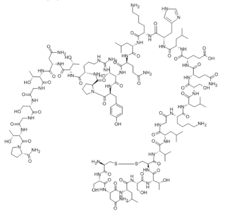
Calcitonin is a 32 amino acid linear polypeptide hormone discovered in 1962 as a hormone postulated to regulate the 'normal level' or 'tone" of calcium in the circulation. It is formed by the proteolytic cleavage of a larger pre-propeptide, which is a product of the CALC1 gene. And it contains s single disulfide bond, which causes the amino terminus to assume the shape of a ring. Alternative splicing of calcitonin pre-mRNA can yield a mRNA encoding calcitonin gene-related peptide; that peptide appears to function in the nervous and vascular systems.
| CAT# | 10-101-08 |
| Product Name | Calcitonin |
| CAS No. | 47931-85-1 |
| Sequence | H-Cys-Ser-Asn-Leu-Ser-Thr-Cys-Val-Leu-Gly-Lys-Leu-Ser-Gln-Glu-Leu-His-Lys-Leu-Gln-Thr-Tyr-Pro-Arg-Thr-Asn-Thr-Gly-Ser-Gly-Thr-Pro-NH2 |
| M.W/Mr. | 3431.9 |
| Molecular Formula | C145H240N44O48S2 |
Function and Work
Initial investigators perfused blood containing high levels of calcium into the thyroid and parathyroid glands of dogs and sheep and discovered the subsequent release of a substance that lowered the blood calcium. The overall effect of calcitonin is to lower the concentration of calcium in the blood when it rides above the normal value. It also lowers the concentration of phosphorus in the blood when levels exceed normal. So calcitonin has been postulated to help regulate normal calcium homeostasis mainly through its biological actions on the bone and kidneys.
Calcitonin works to control calcium and potassium levels. Calcitonin binds to the calcitonin receptors (CTR) on the osteoclast cells to directly inhibit both the number and resorptive capacity of these bone resorbing units, thus decreasing the amount calcium released into the circulation. It does this by inhibiting the activity of the osteoclasts, the cells that break down bone. When the osteoclasts break down bone tissue, the calcium enters the bloodstream. By preventing the breakdown of bone, calcitonin lessens the amount of calcium in the blood. The hormone also seems to decrease the amount of calcium the kidneys can re-absorb, lowing levels further. The calcitonin receptor has been cloned and shown to be member of the seven-transmembrane, G protein-coupled receptor family.
This property of calcitonin has resulted in its use in the treatment of conditions characterized by intensified bone resorption, such as osteoporosis, Paget's disease, and hypercalcemia of malignancy. Calcitonin also inhibits tubular reabsorption of calcium by the kidneys and thus increases the amount of calcium lost in the urine. Although calcitonin is often referred to as being part of the triad of important calcium regulating hormones, along with the 1, 25-dihydroxyvitain D3 and PTH, most of the effects of calcitonin are seen only at pharmacological doses and thus it has been speculated by many that calcitonin may be physiologically unimportant in mammals. Supporting this theory is the fact that there is no significant deficiency syndrome linked solely to the absence of the thyroid, a major source of calcitonin.
There are, however, several studies that suggest calcitonin is important within mammalian physiology. Some of these suggested functions include: acting as an antiresorption agent for the management of bone resorping conditions, a gastrointestinal regulator/satiety controller, a hypocalcemic agent/calciuric hormone, a tumor marker, a regulator of acid-base balance to prevent dehydration, and finally, a regulator of the maternal skeleton during reproduction, just to name a few.
Reference:
Woodrow, Janine P. Calcitonin modulates skeletal mineral loss during lactation through interactions in mammary tissue and directly through osteoclasts in bone. Diss. Memorial University of Newfoundland, 2009.